
- Title
-
Hedgehog signaling and laminin play unique and synergistic roles in muscle development
- Authors
- Peterson, M.T., and Henry, C.A.
- Source
- Full text @ Dev. Dyn.

Somite formation, muscle specification, and muscle differentiation proceed in embryos deficient for both Hh signaling and laminin gamma1. A-J: Side views, anterior left, dorsal top. A-F3: Twenty-two-somite-stage embryos. A-D: DIC images. A1-D1: Stained for β-catenin (white) to outline cells. White arrowheads point to somite boundaries. Note that somite boundaries form in the absence of both laminin and Hh signaling (D, D1). E-F3: In situ hybridizations showing MyoD and myogenin expression in wild-type and lam gamma1 mutant embryos treated with EtOH or CyA. Segmental expression of MyoD and myogenin persists. G-J: Laminin and Hh signaling are required for proper anterior-posterior myosin distribution. Lettered panels: MF20 (light meromyosin) in green. Panels numbered 1 show F310 (myosin light chain) in blue. Panels numbered 2 are histograms that correlate with the images above. The posterior expression domains are indicated by white arrows for EtOH-treated embryos and white arrowheads for CyA-treated embryos. G-G2: EtOH-treated wild-type embryos. MF20 is expressed more posteriorly than F310. H-H2: CyA-treated wild-type embryos. MF20 is not expressed more posteriorly than F310. I-I2: Similar to EtOH-treated wild-type embryos, MF20 is expressed in younger somites than F310 in EtOH-treated lam gamma1 mutant embryos. J-J2: The same shift in myosin chain expression as seen in CyA-treated wild-type embryos is observed in CyA-treated lam gamma1 mutant embryos. K: Graph showing the average posterior limits of MF20 and F310 expression (error bars reflect standard error). L-N3: The lateral-medial pattern of MF20 expression is preserved in CyA-treated embryos. Lettered panels (lateral focal planes) and panels numbered 1 (medial focal planes) are side views, anterior left, dorsal top. Panels numbered 2 and 3 are 3-D reconstructed, transverse views, medial left, lateral right. MF20 expression (white *) is strongest laterally in all embryos (note the lateral peak of MF20 expression in histograms numbered 4). EXPRESSION / LABELING:
PHENOTYPE:
|

Laminin and Hh signaling act synergistically in fast-twitch muscle cell elongation. Side views, anterior left, dorsal top, 48-hpf embryos stained with phalloidin to visualize actin. White arrows point to elongated fast-twitch muscle fibers. White arrowheads points to short, round fast-twitch muscle cells. E-H: Higher-magnification images from a different experiment than A-C. A-C, E-G: Fast-twitch fibers are long in wild-type, lam gamma1 mutant embryos, and CyA-treated wild-type embryos at 48 hpf. D, H: Fast-twitch cells are shorter and round in CyA-treated lam gamma1 mutant embryos. I, J: Three-dimensional projections of dextran-filled cells show the short fast cells in CyA-treated laminin gamma1 mutant embryos. |
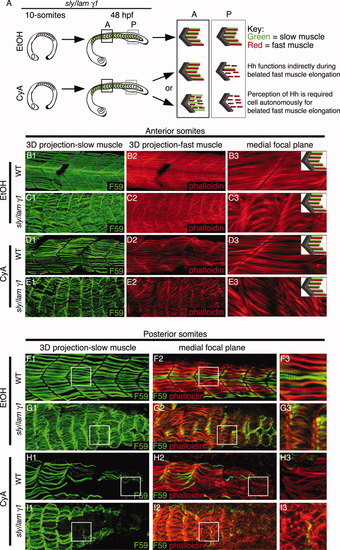
Hh signaling is required indirectly for the recovery of fast-twitch fibers in laminin mutant embryos. B-I: Side views, anterior left, dorsal top, 48-hpf embryos treated with EtOH or CyA at the 10-somite stage and stained with F59 (green) to visualize slow muscle fibers and phalloidin (red) to visualize actin. A: Cartoon describing expected results if Hh signaling is directly or indirectly required. B-E: Anterior somites are shown. Panels numbered 1 are 3-D projections. Panels numbered 3 are higher-magnification views of individual focal planes. Note the presence of slow-twitch fibers and elongated fast fibers in all conditions. F-I: Posterior somites are shown. The boxes in panels numbered 1 and 2 correspond with the higher-magnification medial focal plane shown in panels numbered 3. Slow-twitch fibers are present in EtOH-treated embryos (F, G) but are reduced and/or absent in posterior somites in CyA-treated embryos (H, I). Note that defects in fast fiber elongation correlate with the lack of slow fibers in the posterior of CyA-treated lam gamma1 mutant embryos (I). |