
- Title
-
Use of Phospholipase A(2) for Antigen Retrieval in Zebrafish Whole-Mount Immunohistochemistry
- Authors
- Akhtar, T., Li, J., Olden, T., and Wallace, K.N.
- Source
- Full text @ Zebrafish
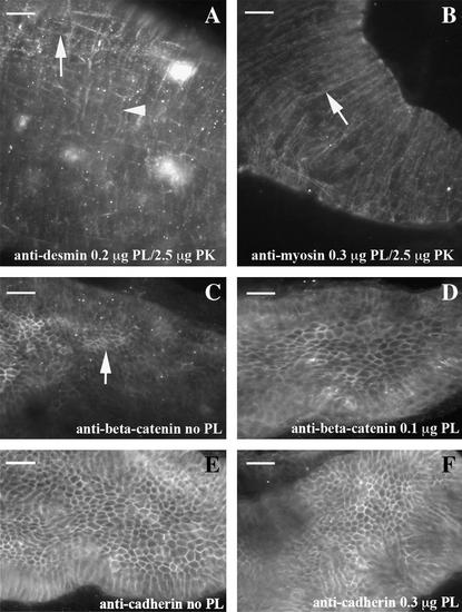
PLA2 retrieves cytoplasmic antigens. Patterns within the dissected intestine are shown with either a combination of PLA2 and proteinase K or PLA2 alone using 96 hpf embryos. Desmin IHC (A) reveals a robust and reproducible pattern with a combination of PLA2 and a low concentration of proteinase K. Both longitudinal muscle (arrow) and circular (arrow head) are observed. Smooth muscle myosin heavy chain IHC (B) shows strong staining within circular muscle (arrow) with a combination of PLA2 and proteinase K. Both beta-catenin (C) and cadherin (E) IHC display part or all of the pattern without antigen retrieval. Addition of an intermediate concentration of PLA2 creates an even pattern of beta-catenin (D), while high levels of PLA2 do not result in distortion of the cadherin pattern (F). In all panels, anterior is left and posterior is right. Scale bar is 20 μM at top left corner. PLA2, phospholipase A2; IHC, immunohistochemistry. |
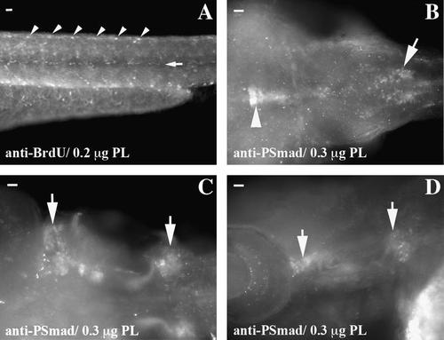
PLA2 retrieves nuclear antigens. Patterns within whole-mount embryos are revealed with PLA2 as the sole enzyme. The pattern of proliferation is identified in 54 hpf embryos injected with BrdU (A). Repeating patterns are observed at somite boundaries (arrowheads) and at somite midline (arrow). Phospho-Smad IHC demonstrates strong nuclear staining in the midbrain (arrowhead B) and hindbrain (arrow B). There is also strong staining in the developing ear (arrows in C and D). In all panels, anterior is left and posterior is right. Scale bar is 20 μM at top left corner. |
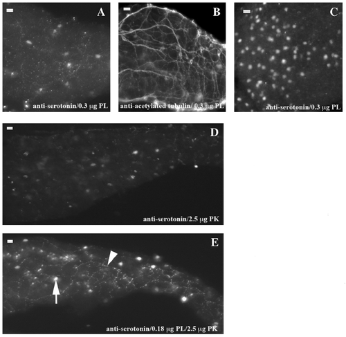
Concentrations of PLA2 are different for nuclear and cytoplasmic serotonin antigen retrieval. High concentrations of PLA2 are required for retrieval of nuclear serotonin within the intestine (A) and pharynx (C) but disrupt serotonin within the axons (A). Overall axon structure is not disrupted at this PLA2 concentration as shown by acetylated tubulin IHC (B). Low concentrations of proteinase K partially permeabilize the tissue (D), while in combination with lower concentrations of PLA2, the full pattern of nuclear (arrow) and axon (arrowhead) staining is present (E). In all panels, anterior is left and posterior is right. Scale bar is 20M at top left corner. |