
|
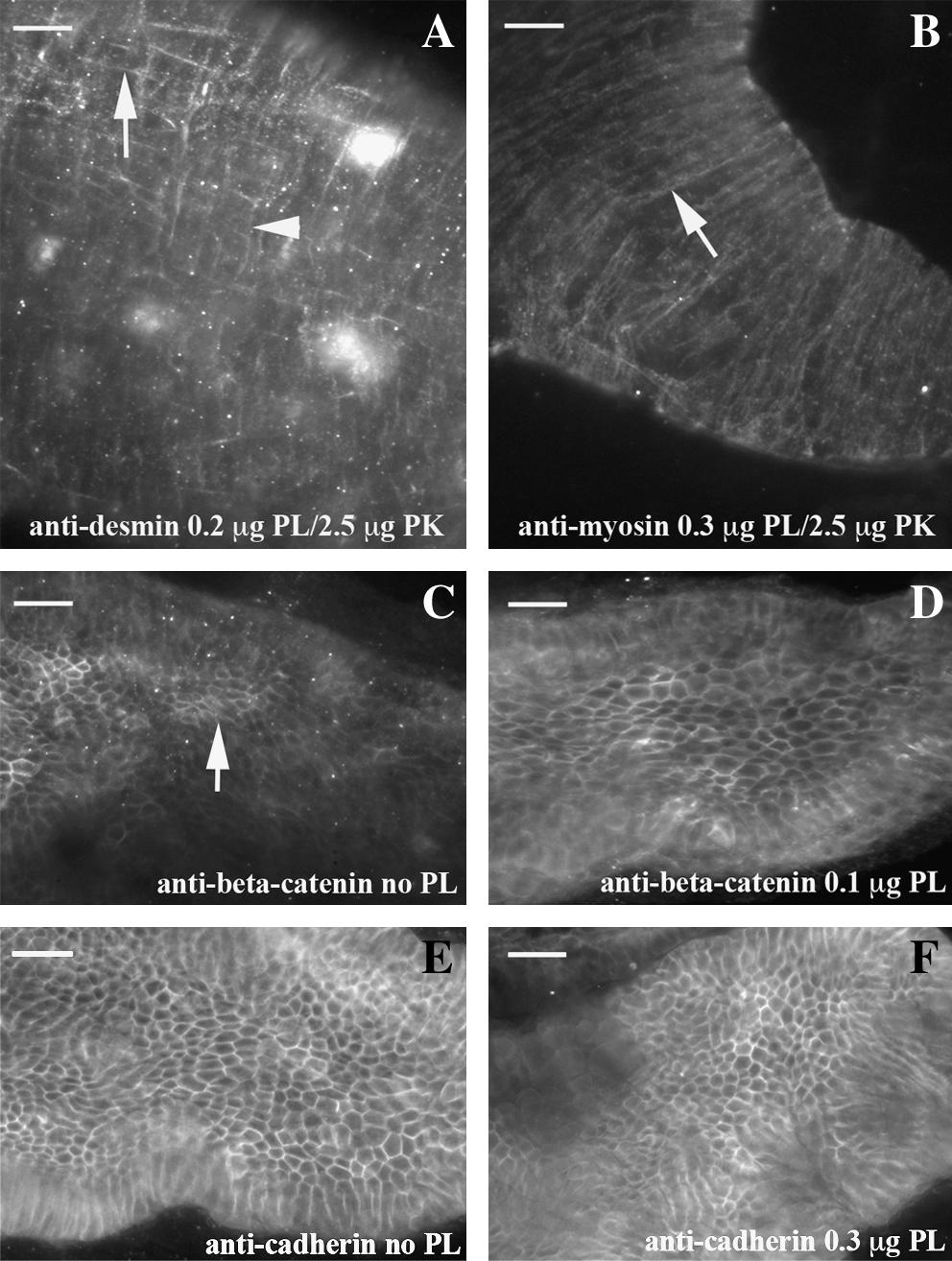
Fig. 1 PLA2 retrieves cytoplasmic antigens. Patterns within the dissected intestine are shown with either a combination of PLA2 and proteinase K or PLA2 alone using 96 hpf embryos. Desmin IHC (A) reveals a robust and reproducible pattern with a combination of PLA2 and a low concentration of proteinase K. Both longitudinal muscle (arrow) and circular (arrow head) are observed. Smooth muscle myosin heavy chain IHC (B) shows strong staining within circular muscle (arrow) with a combination of PLA2 and proteinase K. Both beta-catenin (C) and cadherin (E) IHC display part or all of the pattern without antigen retrieval. Addition of an intermediate concentration of PLA2 creates an even pattern of beta-catenin (D), while high levels of PLA2 do not result in distortion of the cadherin pattern (F). In all panels, anterior is left and posterior is right. Scale bar is 20 μM at top left corner. PLA2, phospholipase A2; IHC, immunohistochemistry.