
Figure 6
- ID
- ZDB-IMAGE-260917-6
- Genes
- Publication
- Chen et al., 2026 - Loss of wbp11 causes multi-system developmental defects: a zebrafish model of VACTERL association
- All Figures
- Figures for Chen et al., 2026
|
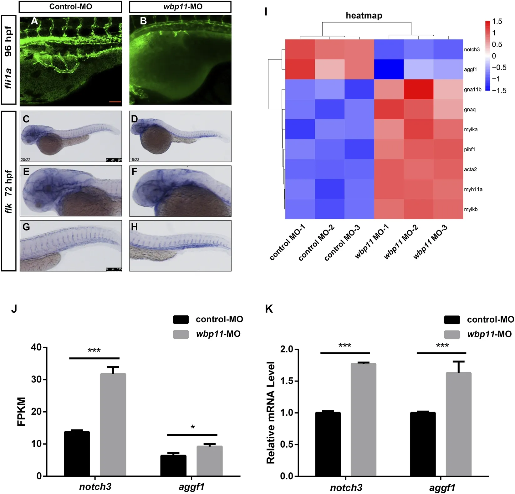
Figure 6 Knockdown of wbp11 causes vascular defects in zebrafish. (A,B) Fluorescence microscopy observation of subintestinal vessels (SIVs) morphology in Tg(fli1a:EGFP) zebrafish larvae at 96 hpf. Red scale bar: 100 μm (applies to (A,B)). (C–H) Whole-mount in situ hybridization of zebrafish larvae at 72 hpf using the flk probe to visualize the vasculature. (C,D) Overview of the entire vascular system. (E,F) Cranial vascular network. (G,H) Truncal vasculature. Black scale bar: 250 μm (applies to (C,D)); black scale bar: 125 μm (applies to (E–H)). (I) Heatmap analysis of vascular-related differentially expressed genes (DEGs). Blue indicates downregulated genes, and red indicates upregulated genes. (J) FPKM values of representative vascular genes notch3 and aggf1 (n = 3; biologically independent experiments, each performed with 50 pooled embryos per group. Data are presented as mean ± SEM. *p < 0.05, ***p < 0.001). (K) qPCR analysis of transcript levels of representative vascular genes notch3 and aggf1 (n = 3; biologically independent experiments, each performed with 50 pooled embryos per group. Data are presented as mean ± SEM. ***p < 0.001).