
Fig. 6
- ID
- ZDB-IMAGE-250819-6
- Genes
- Antibodies
- Publication
- Wu et al., 2025 - Atp7a deficiency induces axonal and myelin developmental defects in zebrafish via ferroptosis
- All Figures
- Figures for Wu et al., 2025
|
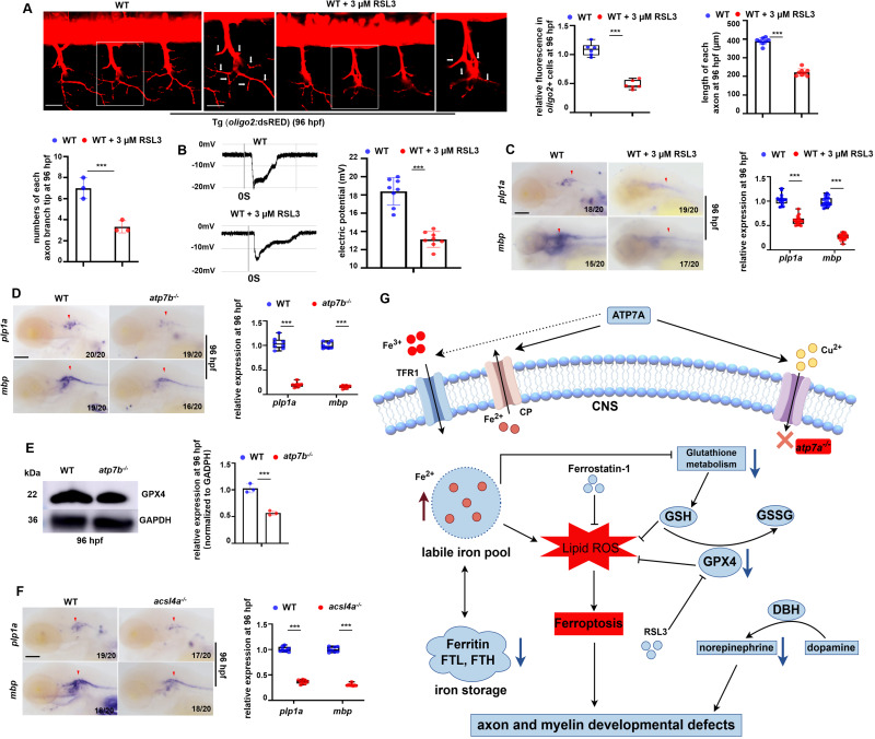
Fig. 6 Effects of unbalanced copper homeostasis and copper non-related ferroptosis on CNS myelin and axon development. (A) oligo2+ cells and the axons in Tg (WT; olig2: dsRED) larvae treated with or without GPX4 inhibitor RSL3 at 96 hpf, and the calculation of the relative fluorescence levels in oligo2+ cell bodies and axons (left), the relative length of axons (right), and the numbers of branch tips in each axon (down)(graphs). (B) Neurons in WT or RSL3 stressed larvae differed in current amplitudes at 96 hpf, with neurons in WT larvae having significantly larger current amplitudes and densities (graphs). (C) Transcriptional expressions of CNS myelin markers mbp and plp1a in WT or RSL3-treated larvae at 96 hpf, and the calculation of the relative expression of mbp and plp1a (box and whisker plots). (D) Transcriptional expressions of CNS myelin markers mbp and plp1a in WT or atp7b−/− larvae at 96 hpf, and the calculations of the relative expressions of mbp and plp1a (box and whisker plots). (E) Western blotting (WB) analysis of the relative protein level of GPX4 in WT or atp7b−/− at 96 hpf, with GADPH as an internal control (graphs). (F) Transcriptional expressions of CNS myelin markers mbp and plp1a in WT or acsl4a−/− larvae at 96 hpf, and the calculations of the relative expressions of mbp and plp1a (box and whisker plots). (G) Working model of atp7a deficiency in inducing developmental defects in axonal and myelin development via ferroptosis in zebrafish. Atp7a functional deficiency leads to damaged iron homeostasis (iron overload) and increased lipid peroxidation, and the subsequent down-regulation of GPX4 and the resulted in ferroptosis in zebrafish larvae, especially in CNS OPCs, and then leads to axon myelin and extension developmental defects and dysfunctional locomotor in zebrafish. C, D, F, lateral view, anterior to the left. Scale bar: 50 μm (A), 10 μm (A, enlarged picture), 100 μm (C, D, F). *P < 0.05, **P < 0.01, ***P < 0.001. NS, not significant.