
|
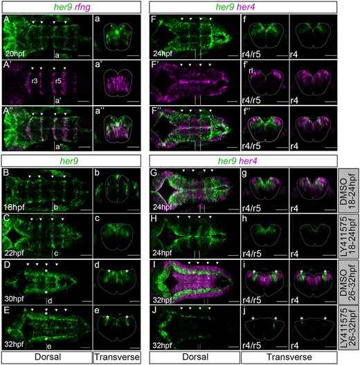
Fig. 1 her9 is temporarily enriched in the hindbrain boundaries independently of Notch at early embryonic stages. (A-J) her9 in situ hybridization with rfng (A′,A″) and her4 (F′,F″,G-J) at the indicated developmental stages. (G-J) Embryos treated with DMSO (G,I) or LY411575 (H,J) (18-24 hpf: n=4/4 DMSO versus n=14/14 LY411575; 26-32 hpf: DMSO n=10/10 versus LY411575 n=8/9). (A-A″,B-E,F-F″,C-J) Dorsal maximal intensity projections of the hindbrain with anterior to the left. (a-a″,b-e,f-f″,g-j) Transverse views of r4/r5 boundary or r4. Arrowheads indicate the position of the hindbrain boundaries. Dotted lines delimitate the contour of the neural tube. Dashed vertical lines indicate the levels of the corresponding transverse projections in other panels. hpf, hours post fertilization; r, rhombomere; rl, rhombic lip. Scale bars: 50 µm.