IMAGE

FIGURE 5

ID
ZDB-IMAGE-250109-298
Genes
Source
Figures for Ercanbrack et al., 2024
Image
Figure Caption

FIGURE 5

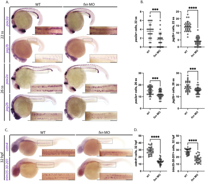
Fxn-deficient zebrafish embryos form reduced multiciliated cell (MCC) progenitors. (A) WISH analysis at the 22 and 26 ss utilizing the probes pax2a and jag2b revealed that the population of MCC progenitors was significantly reduced in fxn morphants compared to wild-type control embryos. Scale bars = 50 um. (B) Unpaired t-test of the corresponding WISH experiments in panel (A). (C) WISH analysis at the 32 hpf time point utilized the probes trim35-30-201 and cetn4, which are expressed by differentiating MCCs, and revealed that MCC numbers were still significantly reduced at this later timepoint. Scale bars = 50 um. Each boxed lateral region in the embryos corresponds to the inset, which shows a dorsal view of that corresponding area. (D) Unpaired t-test of the corresponding WISH experiments in panel (C). ***p < 0.0005, ****p < 0.0001.

Figure Data
Acknowledgments
This image is the copyrighted work of the attributed author or publisher, and ZFIN has permission only to display this image to its users. Additional permissions should be obtained from the applicable author or publisher of the image. Full text @ Front Cell Dev Biol