
Fig. 6
- ID
- ZDB-IMAGE-240725-41
- Genes
- Publication
- Niu et al., 2024 - FBLN2 is associated with Goldenhar syndrome and is essential for cranial neural crest cell development
- All Figures
- Figures for Niu et al., 2024
|
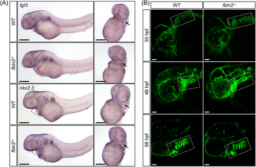
Fig. 6 Normal pharyngeal arch structure in fbln2−/− embryos. (A) WISH at 48 hpf showed no significant difference in the expression of pharyngeal pouch marker fgf3 and nkx2.3, and the pharyngeal pouches were normally generated and segmented in fbln2−/− embryos (magnification factor: 4×, n = 30 per group, bar = 200 μm). The pharyngeal pouches are marked by black dashed rectangles in the lateral view (left) and by black arrows in the ventral view (right). (B) The morphology of pharyngeal arches at 30, 48, and 68 hpf were similar between the wild type and fbln2−/− mutants (magnification factor: 20×, n = 30 per group, bar = 50 μm). The pharyngeal arches are marked by white dashed rectangles. Abbreviation: WT, wild type.