
|
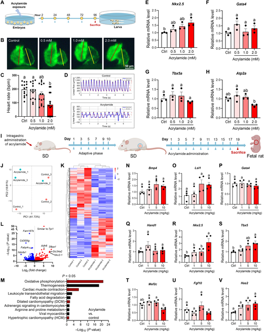
Fig. 7 Acrylamide disturbs heart development during early life stage in both zebrafish embryos and rat embryos. (A) Experimental design: embryos exposed to acrylamide (0.5, 1.0, and 2.0 mM) from 2 to 96 hpf. (B) Representative fluorescence microscopy images of Tg(cmlc2: GFP) zebrafish embryos with green fluorescent protein specifically expressed in the myocardial cells. Embryos at 2 hpf were exposed to acrylamide (0.5, 1.0, and 2.0 mM) for 4 d. (C) Heart rates were measured in zebrafish (2 hpf) treated with acrylamide for 4 d (n = 20 per group). (D) Blood flow dynamics map in control and acrylamide-treated (2.0 mM) zebrafish. (E to H) Relative mRNA expression of cardiac-development-related genes in control and acrylamide-treated zebrafish (n = 3 per group). (I) Experimental design: pregnant rat exposed to acrylamide (1, 5, and 10 mg/kg·bw per day) for 19 d. (J) PCA scores plots based on RNA-seq data of hearts from rat embryos exposed to acrylamide (10 mg/kg·bw per day) (n = 3 per group). (K) Cluster analysis and (L) volcano plot of differentially expressed genes based on RNA-seq analysis. (M) KEGG analysis of RNA-seq data. (N to V) Relative mRNA levels of cardiac-development-related genes in control and acrylamide-treated rat embryos (n = 4 per group). Data are presented as the means ± SEM. Significance was calculated using one-way ANOVA with Tukey’s post hoc test; groups labeled with different letters differed significantly (P < 0.05).