
Fig. 3
- ID
- ZDB-IMAGE-240202-33
- Genes
- Publication
- Nurcombe et al., 2023 - Plexina4 and cell survival in the developing zebrafish hindbrain
- All Figures
- Figures for Nurcombe et al., 2023
|
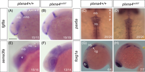
Fig. 3 Development of the hindbrain apparently normal in the absence of Plxna4: Whole mount ISH for hindbrain markers in lateral (A, B, E–H) and dorsal (C, D) views of WT (A, C, E, G) and plxna4 mutant (B, D, F, H) embryos at 22 hpf (A, B) and at 24 hpf (C–H). (A, B) fgf8a marks the midbrain-hindbrain border. (C, D) The transcription factor pax6a. (E, F) sema3fa labels the dorsal midbrain and rhombomere 1 of the hindbrain. (G, H) foxg1a labels the hindbrain and the cranial ganglia. Numbers indicate the number of embryos that exhibit the displayed expression pattern. A: anterior, cg: cranial ganglia, e: eye, fb: forebrain, hb: hindbrain, mb: midbrain, mhb: midbrain-hindbrain boundary, P: posterior, r1: rhombomere 1. Scale bar in A is 250 μm (A, B, E, F), 150 μm (C, D), 300 μm (G, H).