
|
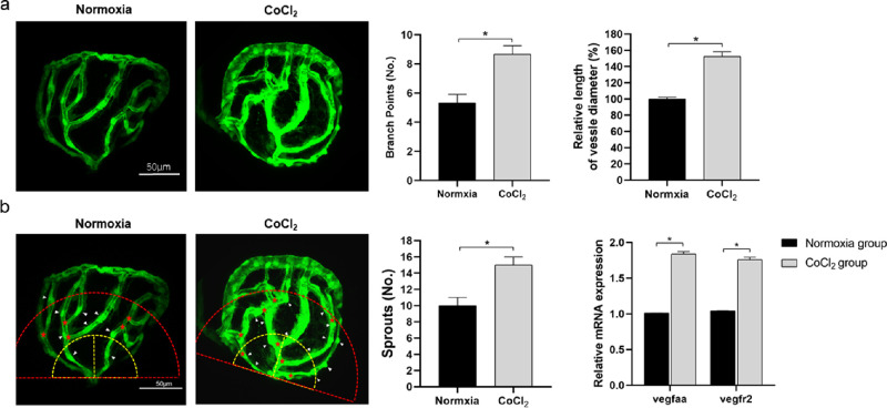
Fig. 9
Comparison of CoCl2-induced retinal neovascularization in zebrafish embryos. (A) With fluorescence excitation, excessive retinal vascularization was shown in 5 mM CoCl2-treated embryos compared with the untreated control at the same developmental stage. (B) The method of counting branch points (red asterisks), vessel sprouts (white arrowheads) within a red semicircle with a radius of 100 µm, and measuring the mean diameter of the retinal vessels proximal to the optic disc (within the yellow semicircle with a radius of 50 µm). Comparison of branch points, vessel diameter and sprouts. mRNA expression of vegfaa and vegfr2 in the retinas of zebrafish embryos. * Significant difference compared with the normoxia group (independent sample t-test, P values < 0.05).