
Fig 13
- ID
- ZDB-IMAGE-221216-36
- Genes
- Publication
- Zhao et al., 2022 - Genetic analysis of activin/inhibin β subunits in zebrafish development and reproduction
- All Figures
- Figures for Zhao et al., 2022
|
Fig 13
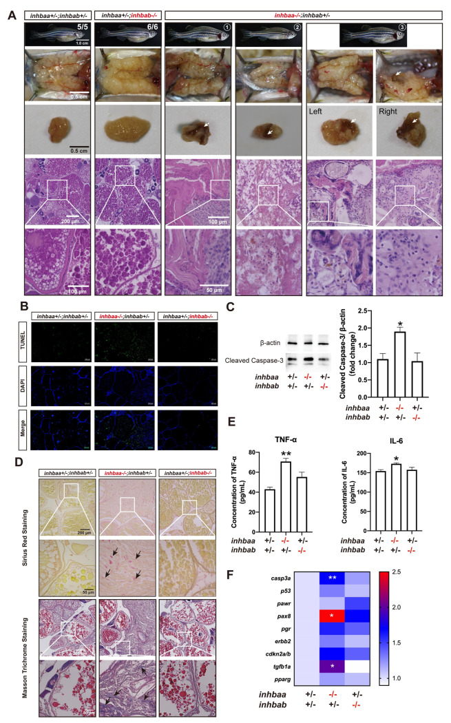
(A) Tumorigenesis in mutant ovaries (>12 mpf). Tumor-like tissues or cysts accumulated in the mutant ovary unilaterally or bilaterally around one-year post-fertilization in all individuals examined (7 in total), and 3 fish contained brownish tissues of different sizes (arrow) (3/7). The fish on the right had the brown tissues on both ovaries. The boxed areas are shown at higher magnification below. (B) TUNEL staining for apoptosis in the ovary. Large areas of somatic cells in the ovary of inhbaa-/- but not inhbab-/- mutant showed strong TUNEL signals at 240 dpf. (C) Western blotting analysis for cleaved Caspase-3 in the ovary of βA single mutants (inhbaa-/-, inhbab-/-) at 240 dpf (n = 3). (D) Sirius red and Masson trichrome staining of the ovarian tissues in different genotypes of the βA mutants at 240 dpf. Arrows indicate staining of collagen fibers. The boxed areas are shown at higher magnification below. (E) Concentrations of two proinflammatory cytokines, TNF-α and IL-6, in the ovary of βA mutants at 240 dpf (n = 3). (F) Expression of genes involved in apoptosis (casp3a and p53), ovarian tumors (pawr, pax8, pgr, erbb2 and cdkn2a/b) and fibrosis (tgfb1a and pparg) in the βA mutants at 240 dpf (*p < 0.05; **p < 0.01; n = 5).