
Fig. 2
- ID
- ZDB-IMAGE-220829-45
- Genes
- Publication
- Sheng et al., 2022 - Sema4C Is Required for Vascular and Primary Motor Neuronal Patterning in Zebrafish
- All Figures
- Figures for Sheng et al., 2022
|
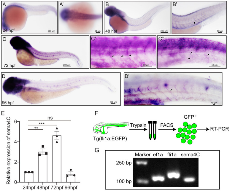
Fig. 2
Expression of sema4C gene in embryonic zebrafish at different stages. (A). Whole mount in situ hybridization (WISH) analysis of sema4C in zebrafish embryos and the magnified image of the head at 24 hpf (A’); (B–D). WISH analysis of sema4C in zebrafish embryos and the magnified images of the trunk at 48 hpf (B’), 72 hpf (C’,C”) and 96 hpf (D,D’), black arrow-heads indicate blood vessels; (E). Expression analysis of sema4C genes in embryonic zebrafish at different stages by QRT-PCR. Triangles, squares, dots and diamonds represent data at different time points respectively. One-way ANOVA, *** p < 0.001, ** p < 0.01, ns, no significance; (F). The procedure of the endothelial cells sorting and RT-PCR; (G). The agarose gel electrophoresis results of RT-PCR on fli1a-EGFP sorted cells.