
Fig. 1
- ID
- ZDB-IMAGE-220811-1
- Genes
- Publication
- Yan et al., 2022 - Irf2bp2a regulates liver development via stabilizing P53 protein in zebrafish
- All Figures
- Figures for Yan et al., 2022
|
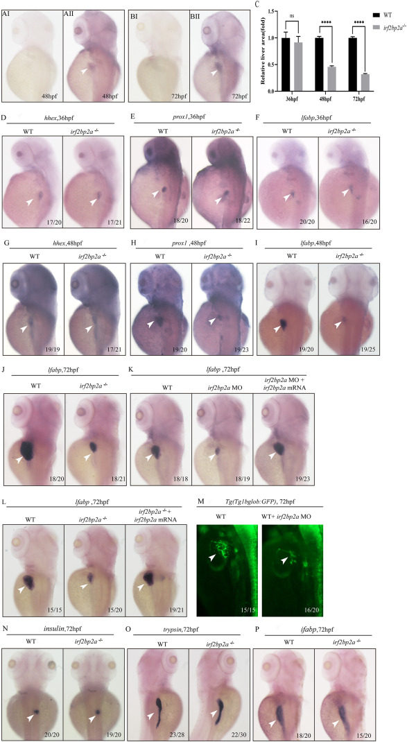
Fig. 1 Fig. 1. Deficiency of zebrafish irf2bp2a led to a reduction of liver size. (A, B) WISH analysis of irf2bp2a expression in zebrafish embryos at 48 hpf and 72 hpf (AII, BII). Embryos incubated with irf2bp2a sense probe are shown as negative controls (AI, BI). (C) The relative liver area measured. The result shown is fold difference compared with the level detected in control embryos. p values are denoted by asterisks. (Student t-test, N ≥ 3. Error bars represent mean ± SD, ns, not significant,****P < 0.0001). (Dsingle bondI) WISH assay of hhex (D, G), prox1 (E, H) and lfabp (F, I) at 36 hpf and 48 hpf, respectively. (M) irf2bp2a MO injection in Tg(Tp1bglob:GFP) line which marks the intrahepatic biliary cells. (J, Nsingle bondP) WISH analysis of lfabp (J), insulin (N), trypsin (O) and ifabp (P) at 72 hpf. (K,L) Irf2bp2a mRNA rescue assays in irf2bp2a−/− embryos and morphant embryos at 72 hpf.
Reprinted from Biochimica et biophysica acta. General subjects, 1866(10), Yan, L., Gao, S., Zhu, J., Zhou, J., Irf2bp2a regulates liver development via stabilizing P53 protein in zebrafish, 130186, Copyright (2022) with permission from Elsevier. Full text @ BBA General Subjects