
Figure 2
- ID
- ZDB-IMAGE-220416-56
- Genes
- Publication
- Huang et al., 2022 - Negative Elongation Factor (NELF) Inhibits Premature Granulocytic Development in Zebrafish
- All Figures
- Figures for Huang et al., 2022
|
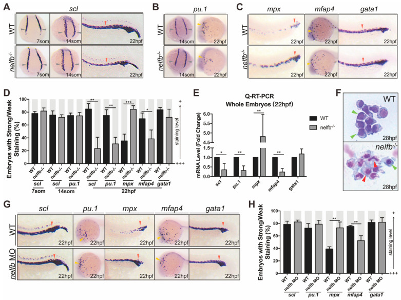
Figure 2 Nelfb deficiency leads to accelerated granulocytic development during primitive hematopoiesis. (A) WISH for scl in WT and nelfb−/− embryos at 7-, 14-somite stages and 22 hpf. (B) WISH for pu.1 in WT and nelfb−/− embryos at 14-somite stage and 22 hpf. (C) WISH for mpx, mfap4, and gata1 in WT and nelfb−/− embryos at 22 hpf. (D) Quantification of WISH results in (A–C) (n = 40–60 embryos per group). (E) Q-RT-PCR analysis of gene expression in nelfb−/− and WT embryos at 22 hpf. Gene expression is normalized to β-actin and presented as fold-change relative to WT. (F) May–Grünwald–Giemsa staining of peripheral blood in WT and nelfb−/− embryos at 28 hpf. Green arrowheads indicate precursors; red arrowheads indicate granulocytes. (G) WISH for scl, pu.1, mpx, mfap4, and gata1 in WT and nelfb morphants at 22 hpf. (H) Quantification of WISH results in (G) (n = 20–40 embryos per group). All results are presented as the mean ± SD from three independent experiments (t test, * for p < 0.05, ** for p < 0.01, *** for p < 0.001). Grey arrowheads, yellow arrowheads, and red arrowheads in (A–C,G), respectively, indicate ICM, ALPM, and PLPM. “+++” and “+” in (D,H) respectively represent strong staining and weak staining. WISH, whole-mount in situ hybridization; ICM, intermediate cell mass; ALPM, anterior lateral plate mesoderm; PLPM, posterior lateral plate mesoderm.