
|
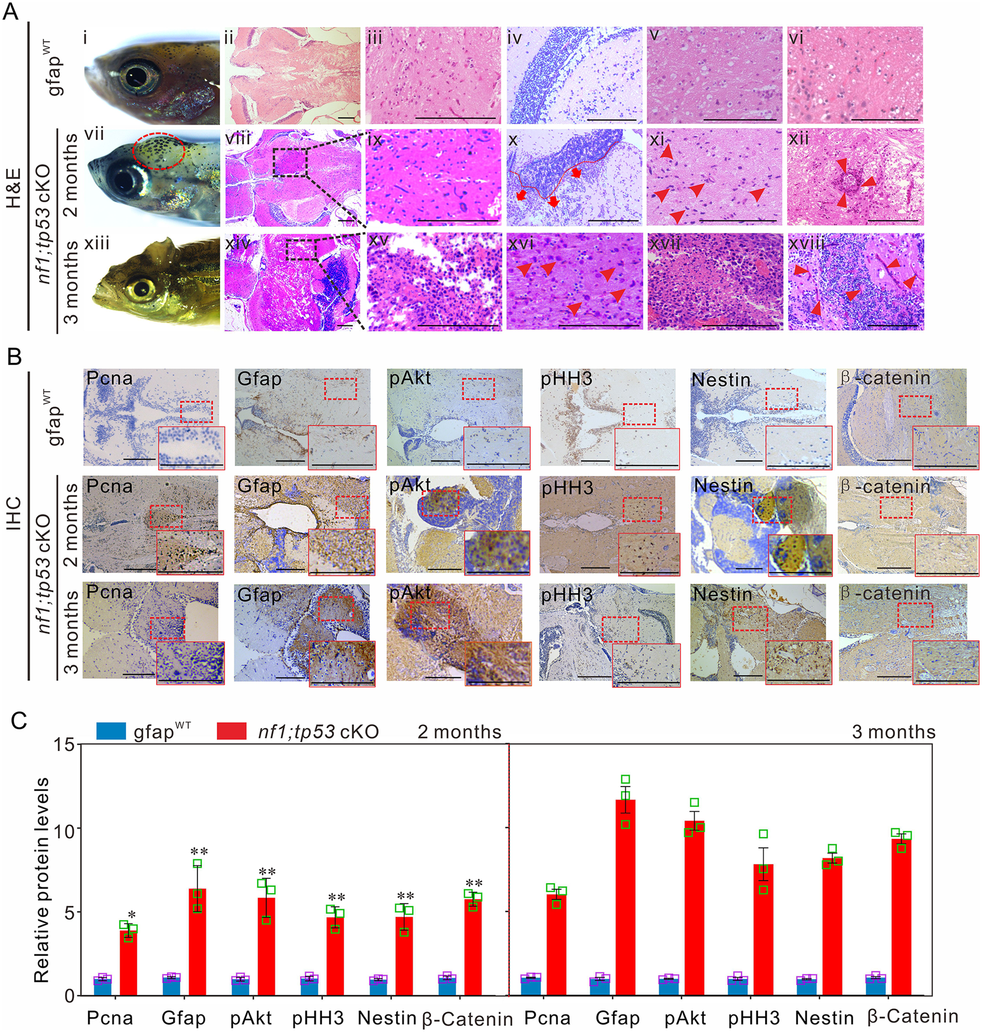
Fig. 4 Tp53 mutation promotes the development of gliomagenesis in zebrafish. (A) Histological examinations of 2-month-old gfapWT and nf1;tp53 cKO fish, and 3-month-old nf1;tp53 cKO fish. (i, vii and xiii) Representative images of the bumps on the heads of nf1;tp53 cKO fish. (ii–vi) Representative images of haematoxylin and eosin (H&E) staining of normal brain tissues of gfapWT fish. Several typical gliomagenic phenotypes, including glioblastoma multiforme (viii and ix), increased numbers of multinucleated glial cells (arrowheads; xi and xvi), necrosis (xiv and xv), frequent vascularity (arrowheads; xii and xviii), enhanced invasive capability (red broken line; x), and the typical gliomatosis phenotype (xvii), were detected in 2- and 3-month-old nf1;tp53 cKO fish. (B) Immunohistochemistry staining was performed to examine the expression of tumour-relevant factors in brain tissues of 2-month-old gfapWT, nf1;tp53 cKO, and 3-month-old nf1;tp53 cKO fish. (C) Quantification of immunohistochemistry staining evaluated the expression of Pcna, Gfap, pAkt, pHH3, Nestin, and β-catenin in 2- or 3-month-old gfapWT and nf1;tp53 cKO fish (n = 3 for each group). Scale bars = 100 μm. Data shown as mean ± SEM. *P < 0.05, **P < 0.01, ***P < 0.001.