IMAGE

Fig. 3

ID
ZDB-IMAGE-210331-15
Genes
Source
Figures for Zhong et al., 2021
Image
Figure Caption

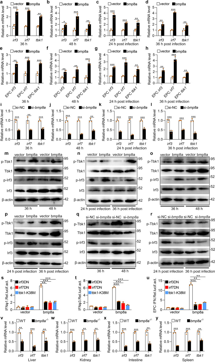
Fig. 3 Bmp8a increases Tbk1–Irf3–Ifn antiviral signaling.

a, b, e, f Expression of irf3, irf7, and tbk1 mRNA after transfected with 2 μg bmp8a or empty vector in ZFL (a, b) or EPC (e, f) cells. The cells were collected at 36 h (a, e) or 48 h (b, f) post-transfection. c, d, g, h Expression of irf3, irf7, and tbk1 mRNA after transfected with 2 μg bmp8a or empty vector in ZFL (c, d) or EPC (g, h) cells for 24 h, followed by infection with GCRV for another 24 h (c, g) or 36 h (d, h). il Expression of irf3, irf7, and tbk1 mRNA after bmp8a knockdown in ZFL cells. The cells were collected at 36 h (i) and 48 h (j) post-knockdown or at 24 h (k) and 36 h (l) post-infected with GCRV. m, o Immunoblot analysis of phosphorylated (p-) Tbk1 and Irf3 after transfected with 2 μg bmp8a or empty vector in ZFL (m) or EPC (o) cells. The cells were collected at 36 or 48 h post-transfection for Immunoblot analysis. n, p Immunoblot analysis of phosphorylated (p-) Tbk1 and Irf3 after transfected with 2 μg bmp8a or empty vector in ZFL (n) or EPC (p) cells for 24 h, followed by infection with GCRV for another 24 or 36 h. q, r Immunoblot analysis of phosphorylated (p-) TBK1 and IRF3 after bmp8a knockdown in ZFL cells. The cells were collected at 36 and 48 h post-knockdown or at 24 and 36 h post-infected with GCRV. su EPC cells were cotransfected with IFN-φ1pro-luc (200 ng, s), IFN-φ3pro-luc (200 ng, t) or EPC IFNpro-luc (200 ng, u), and bmp8a (100 ng) together with each of the dominant negative plasmids including tbk1–K38M (100 ng), irf3DN (100 ng) and irf7DN (100 ng). At 48 h post-transfection, the cells were collected for luciferase assays. Renilla luciferase was used as the internal control. vy Expression of irf3, irf7, and tbk1 mRNA in the liver, kidney, intestine, and spleen from WT or bmp8a−/− zebrafish injected i.p. with 50 µl of GCRV (108TCID50 per ml). The expression of zebrafish actb1 or EPC actin was used as an internal control for the qRT-PCR. Data were from three independent experiments and were analyzed by Student’s t-test (two-tailed) for comparison of two groups or one-way ANOVA followed by Games–Howell post hoc tests for comparison of multiple groups. All data were presented as mean ± SD (**p < 0.01, ***p < 0.001).

Figure Data
Acknowledgments
This image is the copyrighted work of the attributed author or publisher, and ZFIN has permission only to display this image to its users. Additional permissions should be obtained from the applicable author or publisher of the image. Full text @ Commun Biol