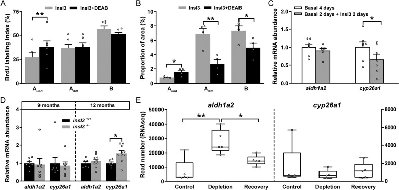
IMAGE

Fig. 5

ID
ZDB-IMAGE-210227-6
Genes
Source
Figures for Crespo et al., 2021
Image
Figure Caption

Fig. 5 Involvement of retinoic acid (RA) signaling in Insl3-induced spermatogenesis.

A, B Evaluation of the proliferation activity (A) and of the proportions of spermatogonia (B) in zebrafish testes cultured for 4 days with 100 ng/mL Insl3, and in the absence or presence of the RA inhibitor DEAB (10 μM). C Ex vivo transcript levels of the RA producing (aldh1a2) and degrading enzymes (cyp26a1) in testis tissue incubated in the absence or presence of 100 ng/mL Insl3. Daldh1a2 and cyp26a1 expression levels in wild-type (insl3+/+) and insl3 knockout (insl3−/−) testes 9 and 12 months post-fertilization. E Read numbers (RNAseq) of RA metabolic enzymes in control, germ cell-depleted (by exposure to the cytostatic agent busulfan51), and testes with recovering (from busulfan) spermatogenesis, as described by Crespo et al.25 (NCBI GEO data set GSE116611). In A, B and E, data are shown as mean ± SEM (AN = 6; BN = 4; EN = 5; *p < 0.05; **p < 0.01), and in C, D as mean fold change ± SEM (C: N = 7; Dinsl3+/+ and insl3−/− 9 months, N = 8 and 7; insl3+/+ and insl3−/− 12 months, N = 6 and 10; *p < 0.05) and expressed relative to the control group (which is set at 1). Aund, type A undifferentiated spermatogonia; Adiff, type A differentiating spermatogonia; B, type B spermatogonia.

Figure Data
Acknowledgments
This image is the copyrighted work of the attributed author or publisher, and ZFIN has permission only to display this image to its users. Additional permissions should be obtained from the applicable author or publisher of the image. Full text @ Commun Biol