
Fig. 5
- ID
- ZDB-IMAGE-200903-8
- Genes
- Publication
- Schumacher et al., 2020 - Integrin α5 and Integrin α4 cooperate to promote endocardial differentiation and heart morphogenesis
- All Figures
- Figures for Schumacher et al., 2020
|
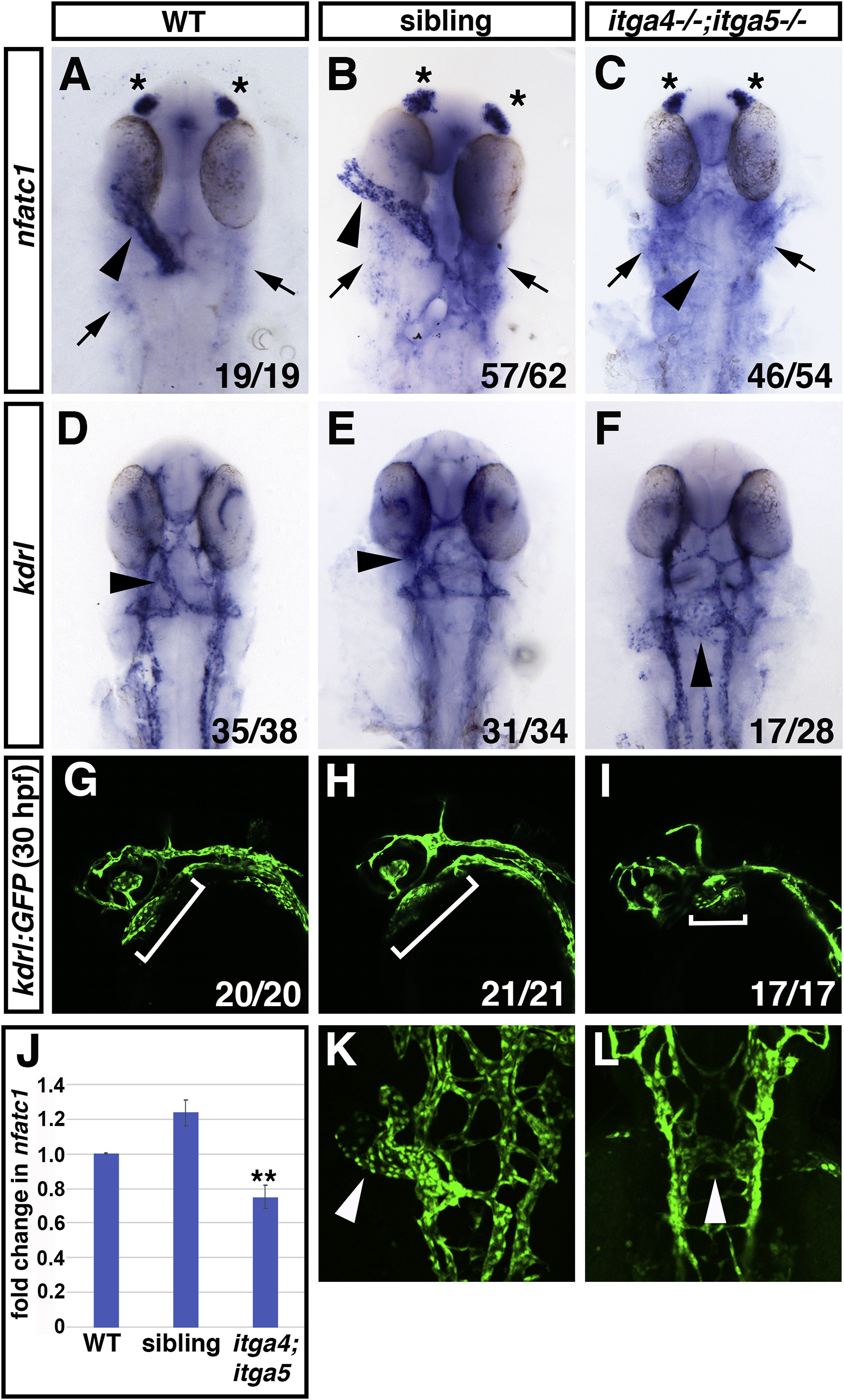
Fig. 5 Fig. 5. itga4;itga5 double mutants have endocardial differentiation and morphology defects. (A–C) nfatc1 expression analyzed by ISH is strongly reduced or absent in the endocardium of itga4;itga5 double mutants compared to siblings and WT embryos. Asterisks indicate nfact1 expression in olfactory placodes. Arrows indicate nfatc1 expression in pharyngeal cells. (D–F) kdrl expression is normal in the endocardium of itga4;itga5 double mutants, but the endocardium does not form a tube in itga4;itga5 double mutants compared to siblings and WT embryos. Dorsal views, arrowheads indicate endocardium. (G-I, K-L) Confocal images of kdrl:GFP expression in endothelial cells reveals that the endocardial tube fails to form in itga4;itga5 double mutants at 30 hpf. Lateral views in G-I, brackets indicate position of endocardial tissue. Dorsal views in K, L, arrowheads indicate endocardial tissue. (J) qPCR for nfatc1 expression at 28 hpf. ∗∗ indicates p < 0.005, comparing itga4;itga5 double mutants to either WT or siblings using Student’s T test. Error bars indicate SEM.
Reprinted from Developmental Biology, 465(1), Schumacher, J.A., Wright, Z.A., Owen, M., Bredemeier, N.O., Sumanas, S., Integrin α5 and Integrin α4 cooperate to promote endocardial differentiation and heart morphogenesis, 46-57, Copyright (2020) with permission from Elsevier. Full text @ Dev. Biol.