
Fig. 7
- ID
- ZDB-IMAGE-200903-6
- Genes
- Publication
- Schumacher et al., 2020 - Integrin α5 and Integrin α4 cooperate to promote endocardial differentiation and heart morphogenesis
- All Figures
- Figures for Schumacher et al., 2020
|
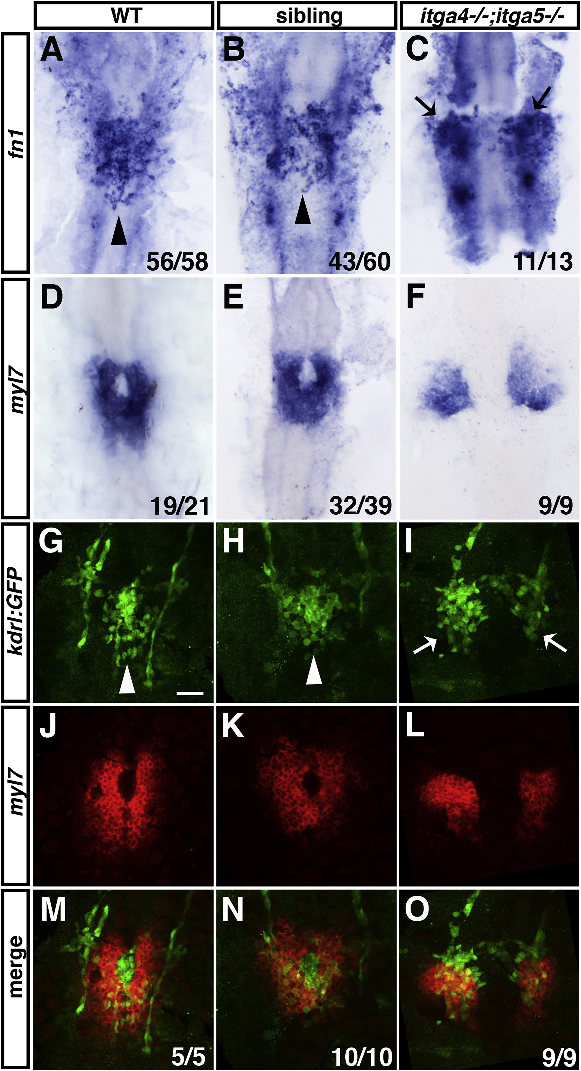
Fig. 7 Fig. 7. Defects in medial migration of endocardial and myocardial cells in itga4;itga5 double mutants. (A–C) fn1 expression in endocardial progenitors is normal but progenitors remain bilateral in itga4;itga5 double mutants compared to sibling and WT embryos. Arrowheads indicate endocardial cells at the midline. Arrows indicate endocardial cells that remain in lateral positions. (D–F) myl7 expression in differentiated myocardial cells is normal but cells remain bilateral in itga4;itga5 double mutants compared to sibling and WT embryos. (G–I) Immunofluorescence of kdrl:GFP expression in the endocardium of WT, sibling embryos, and itga4;itga5 double mutants. White arrowheads indicate endocardial cells at the midline. White arrows indicate endocardial cells in lateral positions. (J–L) Fluorescent ISH for myl7 expression in myocardial cells of WT, sibling embryos, and itga4;itga5 double mutants. (M–O) Overlap of kdrl:GFP and myl7 expression reveals that endocardial and myocardial cells are both located in bilateral regions of the ALPM in itga4;itga5 double mutants. Dorsal views, anterior to the top. All embryos are at 20 somites. Scale bar is 50 μm.
Reprinted from Developmental Biology, 465(1), Schumacher, J.A., Wright, Z.A., Owen, M., Bredemeier, N.O., Sumanas, S., Integrin α5 and Integrin α4 cooperate to promote endocardial differentiation and heart morphogenesis, 46-57, Copyright (2020) with permission from Elsevier. Full text @ Dev. Biol.