
Fig. 4
- ID
- ZDB-IMAGE-200807-21
- Genes
- Publication
- Balogh et al., 2020 - Pseudouridylation defect due to DKC1 and NOP10 mutations causes nephrotic syndrome with cataracts, hearing impairment, and enterocolitis
- All Figures
- Figures for Balogh et al., 2020
|
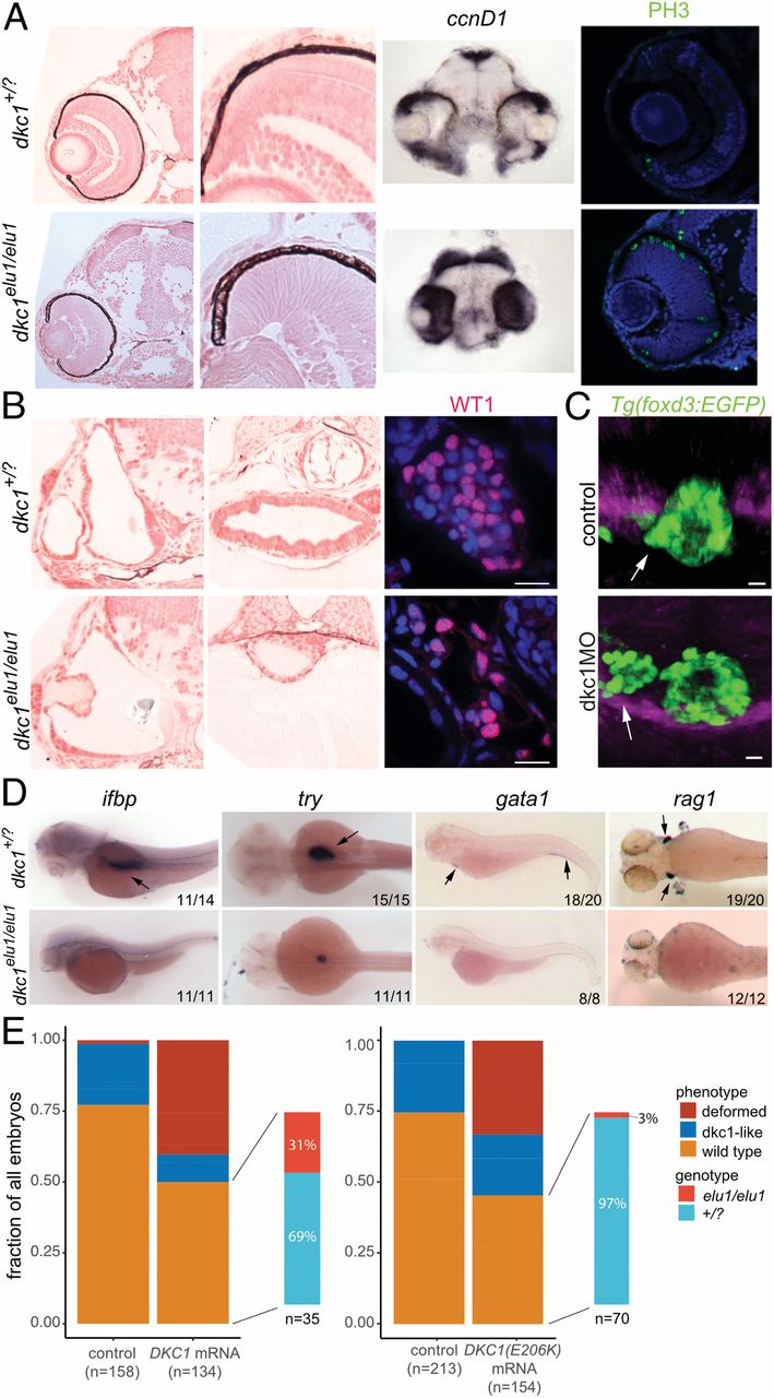
Fig. 4 The phenotype of dkc1elu1/elu1 larvae recapitulates the human phenotype. (A) Histological analysis of dkc1elu1/elu1 mutant larvae shows microphthalmia and cataracts. Both the eyes and the optic tectum of the mutants are abnormal and contain a high prevalence of cells with neuroepithelial character. Expression of cell-cycle markers ccnD1 and PH3 in the retinae and the tecta of 2-dpf and 3-dpf larvae, respectively, can be observed throughout these tissues instead of being restricted to the proliferative regions of the ciliary marginal zone and the mediolateral edges, suggesting a defective cell cycle. (All pictures show coronal sections.) (B) Further histological analysis shows 1) deformed semicircular canals, 2) undifferentiated gut, and 3) hypoplastic pronephros with a reduced number of Wt1-positive podocytes in the mutant animals. (Scale bars: 10 μm.) (C) When Dkc1 function is abrogated in Tg(foxd3:EGFP) animals using a synthetic MO oligo, parapineal migration is impaired, and the pineal–parapineal complex appears immature at 3 dpf. (White arrows denote the parapineal.) (D) Markers of tissue differentiation demonstrate a lack of differentiation in the intestines (ifbp), pancreas (try), and the major blood lineages (gata1 and rag1). (Black arrows denote area of expression.) (E) Injection of 1) human WT DKC1 mRNA resulted in phenotypic rescue of the mutant larvae, as shown by the genotyping of larvae showing a WT phenotype. In contrast, injection of 2) human Glu206Lys DKC1 mRNA elicited a much milder rescue, demonstrating the hypomorphic nature of this allele.