
Fig. 8
- ID
- ZDB-IMAGE-200306-118
- Genes
- Antibodies
- Publication
- Nimura et al., 2019 - Role of Reelin in cell positioning in the cerebellum and the cerebellum-like structure in zebrafish
- All Figures
- Figures for Nimura et al., 2019
|
Fig. 8
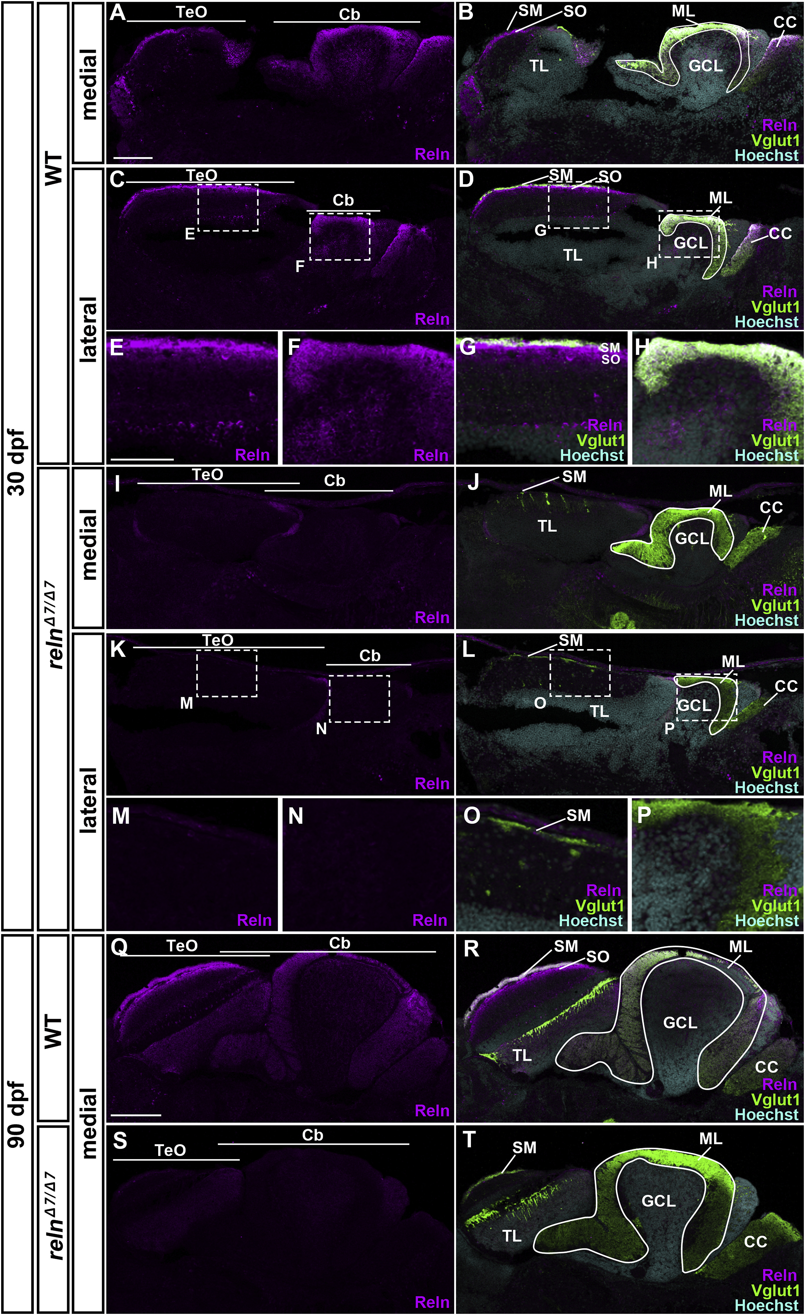
Localization of Reln protein in the tectum and the cerebellum. Medial and lateral sagittal sections of the brain from 30-dpf (A–P) and 90-dpf (Q–T) WT and relnΔ7/Δ7 mutant zebrafish were stained with anti-Reln (magenta), anti-Vglut1 (green), and Hoechst (cyan). Three fish for 30-dpf and one fish for 90-dpf WT or relnΔ7/Δ7 mutant fish were analyzed. Typical images are shown. (E-H, M-P) High magnification images of the boxes in C, D, K, and L. In WT, Reln protein was detected strongly in the SM and relatively weakly in the SO in the TeO. Reln was also detected strongly in the ML in the Cb. Weak Reln signals were also detected in the GCs in the TL and the GCL. These signals were absent in relnΔ7/Δ7mutants. CC, crista cerebellaris. The other abbreviations are described in the legend for Fig. 1. Scale bars: 100 μm in A (applies to A-D, I-L); 50 μm in E (applies to E-H, M-P); 200 μm in Q (applies to Q-T).
Reprinted from Developmental Biology, 455(2), Nimura, T., Itoh, T., Hagio, H., Hayashi, T., Di Donato, V., Takeuchi, M., Itoh, T., Inoguchi, F., Sato, Y., Yamamoto, N., Katsuyama, Y., Del Bene, F., Shimizu, T., Hibi, M., Role of Reelin in cell positioning in the cerebellum and the cerebellum-like structure in zebrafish, 393-408, Copyright (2019) with permission from Elsevier. Full text @ Dev. Biol.