
Fig. S3
- ID
- ZDB-IMAGE-200121-9
- Genes
- Publication
- Yang et al., 2019 - RNA 5-Methylcytosine Facilitates the Maternal-to-Zygotic Transition by Preventing Maternal mRNA Decay
- All Figures
- Figures for Yang et al., 2019
|
Fig. S3
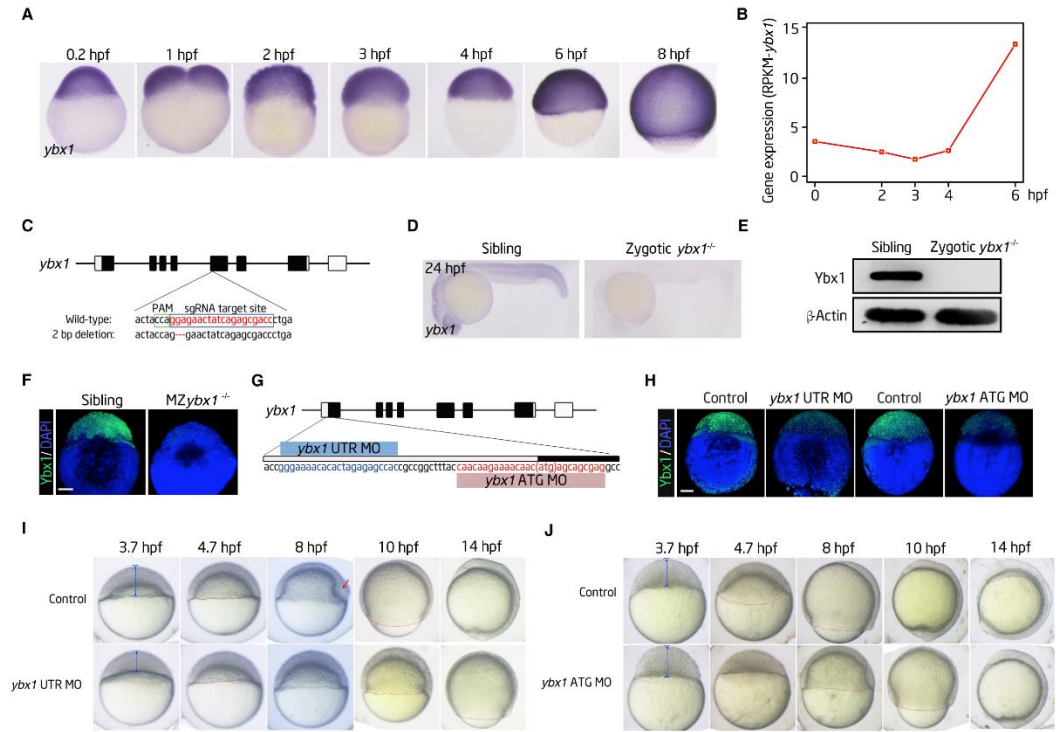
Generation and Characterization of Ybx1 Mutant Zebrafish and Phenotypes of Ybx1 MO-injected Embryos. Related to Figure 3. (A) In situ hybridization results revealing the expression profile of ybx1 mRNA. (B) RNA-Seq results revealing the expression profile of ybx1 mRNA. The expression levels were normalized by the RPKM of gapdh in the corresponding stages. (C) Graphical representation of the target site in the fifth exon of ybx1 used for the design of mutants through a CRISPR/Cas9 strategy. The lower panel shows the 2 bp insertion in the ybx1 target site. (D) Whole mount in situ hybridization analysis results showing the lack of expression of ybx1 in the ybx1 mutant. (E and F) Western blotting (E) and immunofluorescence (F) results showing that Ybx1 protein was undetectable in the homozygous mutants. -Actin served as a protein loading control. Scale bar: 200 m. (G) Design of the ybx1 MO that targeted the 5’UTR (UTR MO) or the start codon (ATG MO) of ybx1 mRNA.