
Fig. 3
- ID
- ZDB-IMAGE-180912-13
- Genes
- Antibodies
- Publication
- Kinoshita et al., 2018 - Functional roles of the Ripply-mediated suppression of segmentation gene expression at the anterior presomitic mesoderm in zebrafish
- All Figures
- Figures for Kinoshita et al., 2018
|
Fig. 3
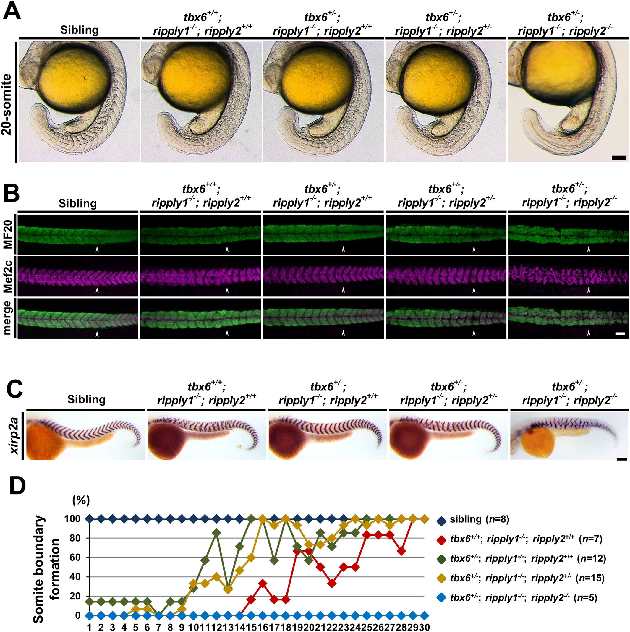
Recovery of the somite boundary in tbx6+/−; ripply1−/− embryos depends on ripply2. (A) Lateral views of sibling, tbx6+/+; ripply1−/−; ripply2+/+, tbx6+/−; ripply1−/−; ripply2+/+, tbx6+/−; ripply1−/−; ripply2+/−, and tbx6+/−; ripply1−/−; ripply2−/− embryos at the 20-somite stage. These embryos were obtained by intercross between tbx6+/−; ripply1+/−; ripply2+/− triple heterozygous mutants. (B) Immunostaining of the myotomes of sibling, tbx6+/+; ripply1−/−; ripply2+/+, tbx6+/−; ripply1−/−; ripply2+/+, and tbx6+/−; ripply1−/−; ripply2+/− embryos at 36 hpf using MF20 and anti-Mef2c antibodies. The arrowhead shows the posterior end of the yolk extension. Anterior is to the left. (C) Expression patterns of a segment boundary marker, xirp2a, in sibling, tbx6+/+; ripply1−/−; ripply2+/+, tbx6+/−; ripply1−/−; ripply2+/+, tbx6+/−; ripply1−/−; ripply2+/−, and tbx6+/−; ripply1−/−; ripply2−/− embryos at 36 hpf. (D) Distribution of somite boundary formation in tbx6+/+; ripply1−/−; ripply2+/+, tbx6+/−; ripply1−/−; ripply2+/+, tbx6+/−; ripply1−/−; ripply2+/−, and tbx6+/−; ripply1−/−; ripply2−/− embryos at 36 hpf. Scale bars in A, B, and C are 100 μm.
Reprinted from Mechanisms of Development, 152, Kinoshita, H., Ohgane, N., Fujino, Y., Yabe, T., Ovara, H., Yokota, D., Izuka, A., Kage, D., Yamasu, K., Takada, S., Kawamura, A., Functional roles of the Ripply-mediated suppression of segmentation gene expression at the anterior presomitic mesoderm in zebrafish, 21-31, Copyright (2018) with permission from Elsevier. Full text @ Mech. Dev.