
Fig. 5
- ID
- ZDB-IMAGE-180705-11
- Genes
- Publication
- Liu et al., 2018 - Lipoprotein lipase regulates hematopoietic stem progenitor cell maintenance through DHA supply
- All Figures
- Figures for Liu et al., 2018
|
Fig. 5
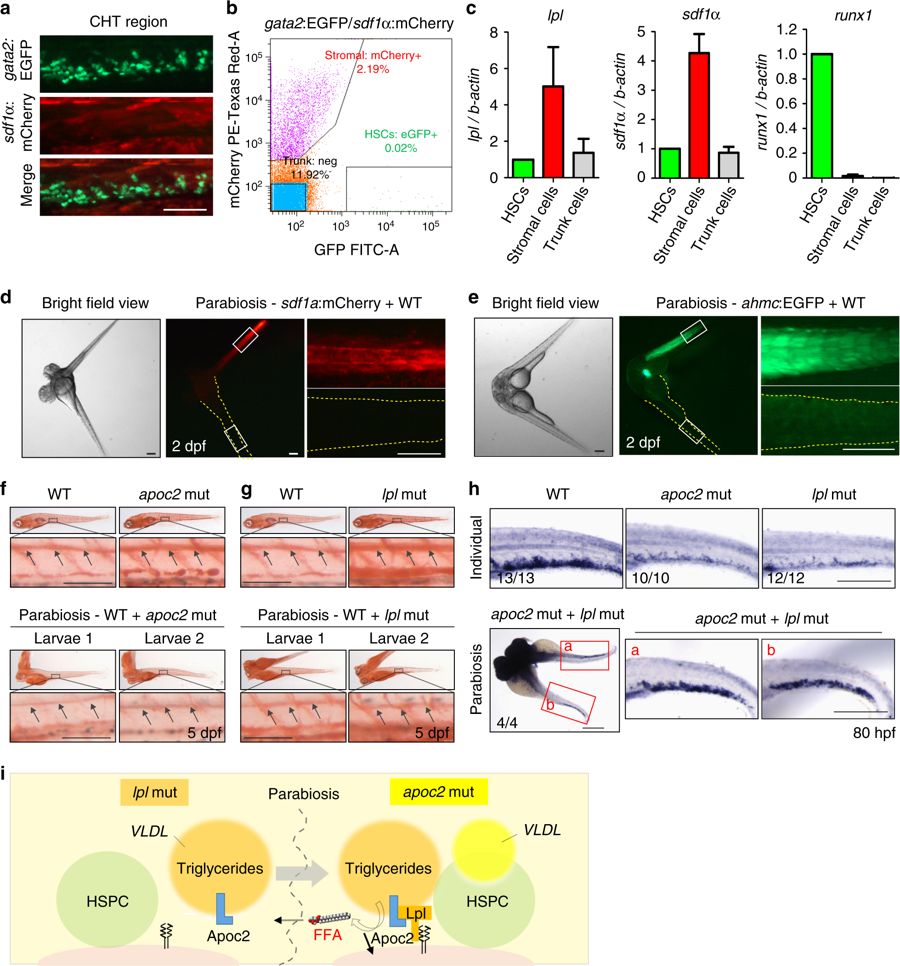
Parabiosis of apoc2 and lpl mutants rescues defective hematopoiesis. a The CHT region of gata2:EGFP, sdf1a:mCherry double-positive embryos at 2.5 dpf. b Flow cytometry of gata2:EGFP and sdf1a:mCherry positive cells isolated from the CHT region. c RT-qPCR analysis of FACS-sorted gata2:EGFP and sdf1a:mCherry positive cells, using lpl, runx1 and sdf1α primers. Mean ± SD of two independent experiments. d, e Parabiosis of a sdf1α:mCherry or a ahmc:EGFP with a WT embryo. Right-hand panels are enlarged segments showed in white quadrangles in left-hand panels. Yellow dashed lines trace WT embryos’ boundaries. f, g Rescue of hyperlipidemia in apoc2 or lpl mutants by parabiosis with WT embryos. Upper panels: two separated embryos (WT and apoc2 or lpl). Lower panels: larva 1 and larva 2 from a parabiosis pair (WT with apoc2 or WT with lpl). Black arrows point to ORO staining in the lumen of blood vessels. h In situ hybridization with runx1/cmyb probe in WT, individual apoc2 and lpl mutants, and parabiotic apoc2 and lpl mutants at 80 hpf. Scale bars, 50 μm in f, g, and 200 μm in a, d, e and h. i Diagram of lipoprotein metabolism in parabiotic lpl and apoc2 mutants. In the lpl mutant, no Lpl is expressed. However, VLDL secreted by the lpl mutant (orange) delivers Apoc2 through the shared circulation to the apoc2 mutant, in which Lpl is expressed but its own VLDL (yellow) contains no Apoc2. VLDL from the lpl mutant compensates lack of Apoc2 in the apoc2 mutant and the reconstituted Apoc2/Lpl catalyzes hydrolysis of TG to release FFAs into the shared circulation, which in turn rescue the hematopoiesis defect in both mutants