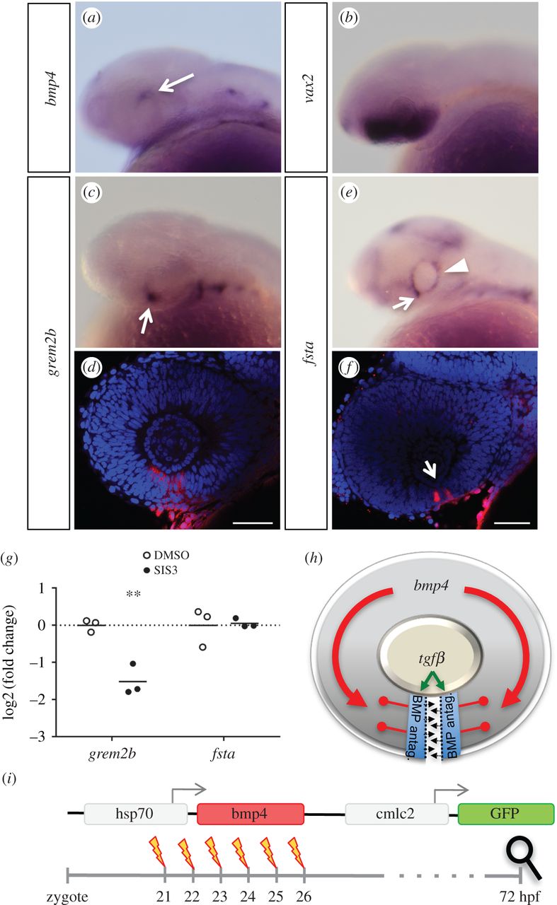
Fig. 3
BMP antagonists grem2b and fsta are expressed in the optic fissure. WMISHs were performed at 30 hpf and are shown in lateral view. (a) bmp4 is expressed in the dorsal optic cup (arrow). (b) Expression of vax2 in the ventral retina. (c,d) Expression of grem2b in the optic cup is restricted to the optic fissure (arrow). (d) Confocal view of grem2b expression (red) with DAPI counterstaining (blue). (e,f) fsta is expressed in the optic fissure (arrows), as well as the ciliary marginal zone (CMZ, arrowhead). (f) Confocal view of fsta expression (red) with DAPI counterstaining (blue). (g) Expression analysis of gremlin and follistatin by quantitative PCR, differential expression in heads of SIS3-treated embryos (30 hpf) as represented by the log2(fold change) of individual samples. Embryos were treated from 24 hpf onward, controls were treated with DMSO. Material from three individuals was pooled for one sample; n = 3, horizontal bars represent the arithmetic mean. p-Values for grem2b and fsta, 4.3 × 10⁻³ and 0.877, respectively. (h) Model of the proposed role of TGFβ and BMP antagonism during optic fissure fusion. TGFβ signalling domains in the optic fissure margins are shielded from BMP by induced BMP antagonists. (i) Scheme of a heat shock inducible BMP construct used to create the transgenic line tg(hsp70:bmp4, cmlc2:GFP). GFP expressed under the cardiac cmlc2 promoter serves as transgenesis marker. Experimental procedure using heat shocks at different time points between 21 and 26 hpf to induce bmp4 expression, aiming at disrupting optic fissure fusion. Analysis of phenotypes was scheduled for 3 dpf.

