
Fig. 2
|
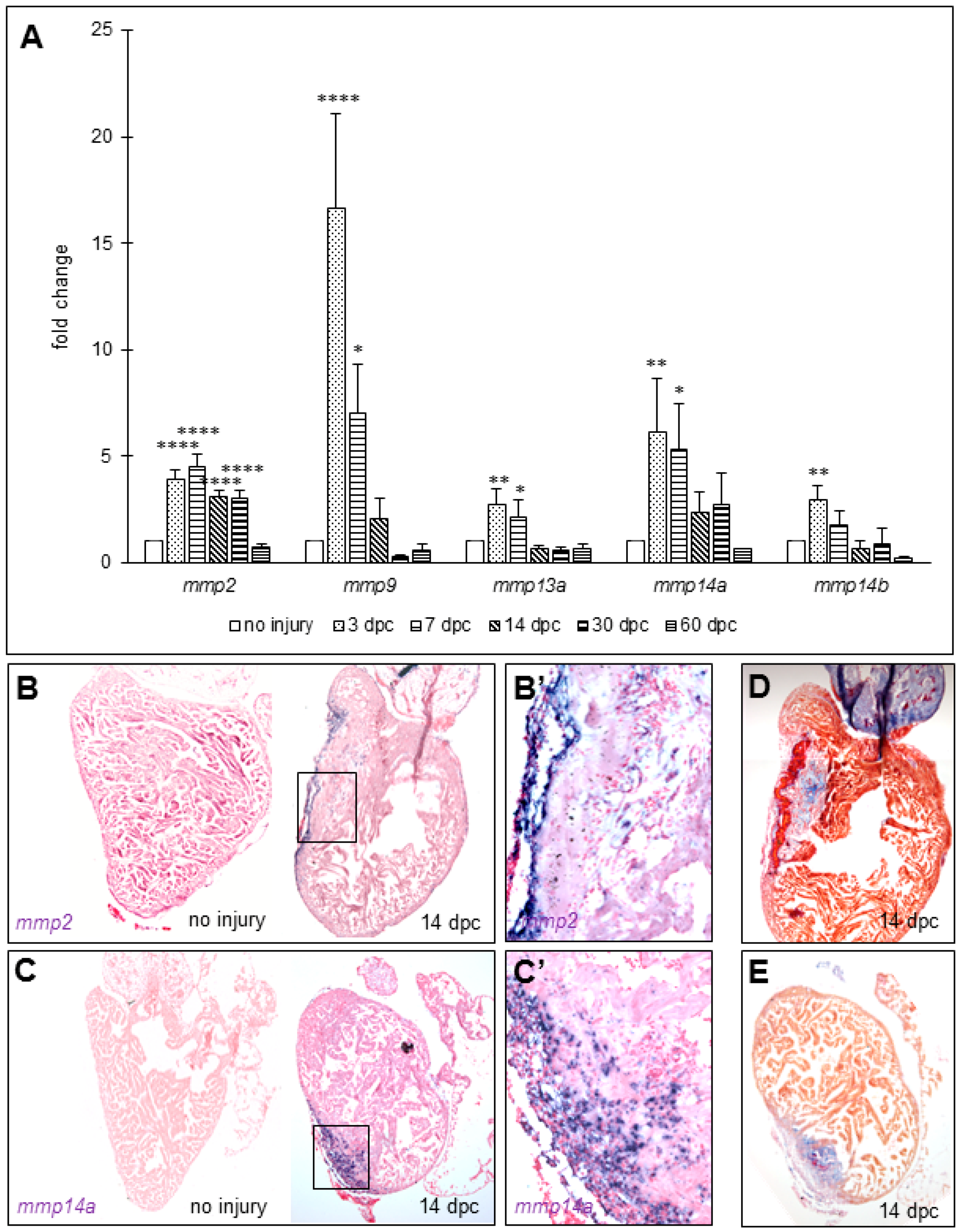
Fig. 2 MMPs transcripts increase in zebrafish ventricles after cryoinjury. (A) RT-quantitative PCR for mmp2, mmp9, mmp13a, mmp14a, and mmp14b in ventricles at several time points after cryoinjury (six to eight ventricles per time point). Significant difference with uninjured hearts: **** p < 0.0001, ** p < 0.01, * p < 0.05; (B,C) in situ hybridization in uninjured and 14 dpc hearts of mmp2 (n = 1 and 2, respectively) and mmp14a (n = 2 and 2, respectively) transcripts respectively; (B’,C’) Magnification of the boxes in (B,C), respectively; (D,E) AFOG staining on consecutive sections of 14 dpc hearts shown in (B,C) respectively for comparison with the wound area (fibrin in red, collagen in blue, and counterstaining in orange).