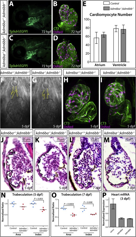
Fig. 4 Kdm6ba and kdm6bb redundantly promote cardiac trabeculation. (A, C) Fluorescent imaged whole mount 72 hpf embryos showing the aortic arch arteries of control and kdm6b-deficient Tg(kdrl:EGFP) animals. (B, D) Confocal heart images of whole mount 72 hpf Tg(myl7:DsRed-nuc)larvae antibody stained for DsRed (red, cardiomyocyte nuclei) and cardiac troponin (CT3, heart muscle). Control and combined kdm6ba/kdm6bb-deficient larvae are shown. (E) Bar graphs comparing the absolute number of atrial and ventricular cardiomyocytes between 72 hpf control (kdm6ba+/-; kdm6bb+/- and kdm6ba+/+; kdm6bb+/-) and kdm6ba-/-; kdm6bb-/- larvae. Error bars represent one standard deviation (n=6 control and 7 kdm6ba/bb-deficient fish from two clutches). (F, G) DIC microscopy images showing the OFT and ventricle of control and kdm6b-deficient embryos at 5 dpf. Dashed yellow lines outline the OFT valves. (H, I) Whole mount confocal imaged hearts of immunostained 5 dpf control and kdm6ba-/-; kdm6bb-/- larvae. These Tg(myl7:DsRed-nuc) fish are stained with anti-DsRed (red, cardiomyocyte nuclei) and anti-cardiac troponin (CT3, green) antibodies. (J-M) H&E stained sagittal sections through the heart of 5 and 7 dpf control and kdm6ba-/-; kdm6bb-/- larvae. Solid lines outline the trabeculae and dashed yellow lines indicate the AVC valves. (N, O) Scatterplot graphs showing the normalized outer curvature ventricular sectional area and trabeculation extent (“index”) of 5 and 7 dpf control compared to clutch mate kdm6ba-/-; kdm6bb-/- larvae. Each data point represents one animal. P-values are from Student's two-tailed t-tests. (P) Bar graph of qRT-PCR data comparing kdm6ba and kdm6bb to vmhcl transcript levels using three pools of isolated 3 dpf zebrafish embryonic hearts (normalized to vmhcl levels). Error bars are one standard deviation.
Reprinted from Developmental Biology, 426(1), Akerberg, A.A., Henner, A., Stewart, S., Stankunas, K., Histone demethylases Kdm6ba and Kdm6bb redundantly promote cardiomyocyte proliferation during zebrafish heart ventricle maturation, 84-96, Copyright (2017) with permission from Elsevier. Full text @ Dev. Biol.

