
Fig. 4
- ID
- ZDB-IMAGE-170825-34
- Genes
- Antibodies
- Publication
- Wehner et al., 2017 - Wnt signaling controls pro-regenerative Collagen XII in functional spinal cord regeneration in zebrafish
- All Figures
- Figures for Wehner et al., 2017
|
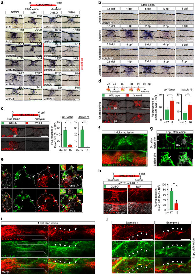
Fig. 4
Wnt/β-catenin signaling controls col12a1a/b transcription and Col XII deposition in a spinal lesion site. a Wnt/β-catenin pathway inhibition (IWR-1) results in lower expression of select collagen genes (red box) in the lesion site at 1 dpl, determined by in situ hybridization. b col12a1a and col12a1b expression is transiently upregulated in the lesion site during regeneration. c Inhibition of Wnt/β-catenin signaling (IWR-1) interferes with col12a1a/b expression in the lesion site (t-test: ***P < 0.001). d wnt8 overexpression upregulates col12a1a/b expression in the lesion site in hs:wnt8 transgenic fish (t-test: *P < 0.05, **P < 0.01). Signal was underdeveloped to optimize detection of differences in signal strength. e A subpopulation of col12a1a or col12a1b-expressing cells (red) in the lesion site co-labels with transcripts of the 6xTCF:dGFP Wnt reporter (green, arrows). f,g Anti-Col XII immunoreactivity is increased in the lesion site (arrows) compared to adjacent unlesioned trunk tissue at 1 dpl. Shown is a confocal image of a whole mount (f, confocal depth was limited to spinal cord) and a transversal section though the lesion site g,h Inhibition of Wnt/β-catenin signaling (IWR-1) interferes with Col XII deposition in the lesion site (t-test: **P < 0.01). Note continuity of tissue in IWR-1-treated animals as shown by re-epithelialization of the lesion site by basal keratinocytes in krtt1c19e:EGFP transgenic fish. i,j Double immunolabelling of axons (anti-acetylated Tubulin+) and Col XII shows Col XII matrix deposition in the lesion site through which regenerating axons grow i. Shown is a maximum intensity projection in i with inset higher magnification; the confocal depth was limited to spinal cord. Arrowheads point to regenerating axonal fascicle in close apposition to Col XII immunoreactivity (also see Supplementary Movie 5). Note, that the trajectory of axonal fascicles appear to follow longitudinal fibres of Col XII immunoreactive ECM material in the lesion site (j, arrowheads). a–j Views are lateral (a–d,f,h–j; dorsal is up, rostral is left) or transversal (e,g; dorsal is up). BF: brightfield. Scale bars: 100 µm a–d,f,h, 50 µm e,g,i, 25 µm j. Error bars indicate s.e.m