
IMAGE
Fig. 7
- ID
- ZDB-IMAGE-170606-25
- Genes
- Antibodies
- Publication
- Naylor et al., 2017 - Wnt8a expands the pool of embryonic kidney progenitors in zebrafish
- All Figures
- Figures for Naylor et al., 2017
Image
|
Figure Caption
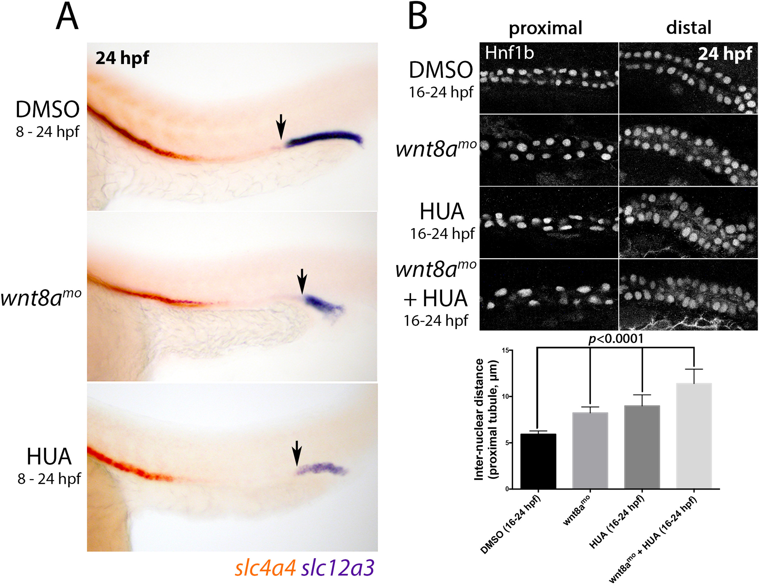
Fig. 7
Effect of wnt8a knockdown on inter-nuclear distance. A) Lateral views of 24 hpf embryos stained for slc4a4 (red) and slc12a3 (purple). Black arrows identify the anterior end of the slc12a3+ DL segment. B) Panels show lateral views of the pronephric tubule (proximal left, distal right) stained for Hnf1b to identify the nucleus of renal epithelial cells in response to wnt8a knockdown and/or HUA treatment. Histogram shows the average inter-nuclear distances of cells in the proximal region of the pronephros for each treatment.
Figure Data
Acknowledgments
This image is the copyrighted work of the attributed author or publisher, and
ZFIN has permission only to display this image to its users.
Additional permissions should be obtained from the applicable author or publisher of the image.
Reprinted from Developmental Biology, 425(2), Naylor, R.W., Han, H.I., Hukriede, N.A., Davidson, A.J., Wnt8a expands the pool of embryonic kidney progenitors in zebrafish, 130-141, Copyright (2017) with permission from Elsevier. Full text @ Dev. Biol.