
Fig. 2
- ID
- ZDB-IMAGE-160729-33
- Genes
- Publication
- Xu et al., 2014 - Maternal Vsx1 plays an essential role in regulating prechordal mesendoderm and forebrain formation in zebrafish
- All Figures
- Figures for Xu et al., 2014
|
Fig. 2
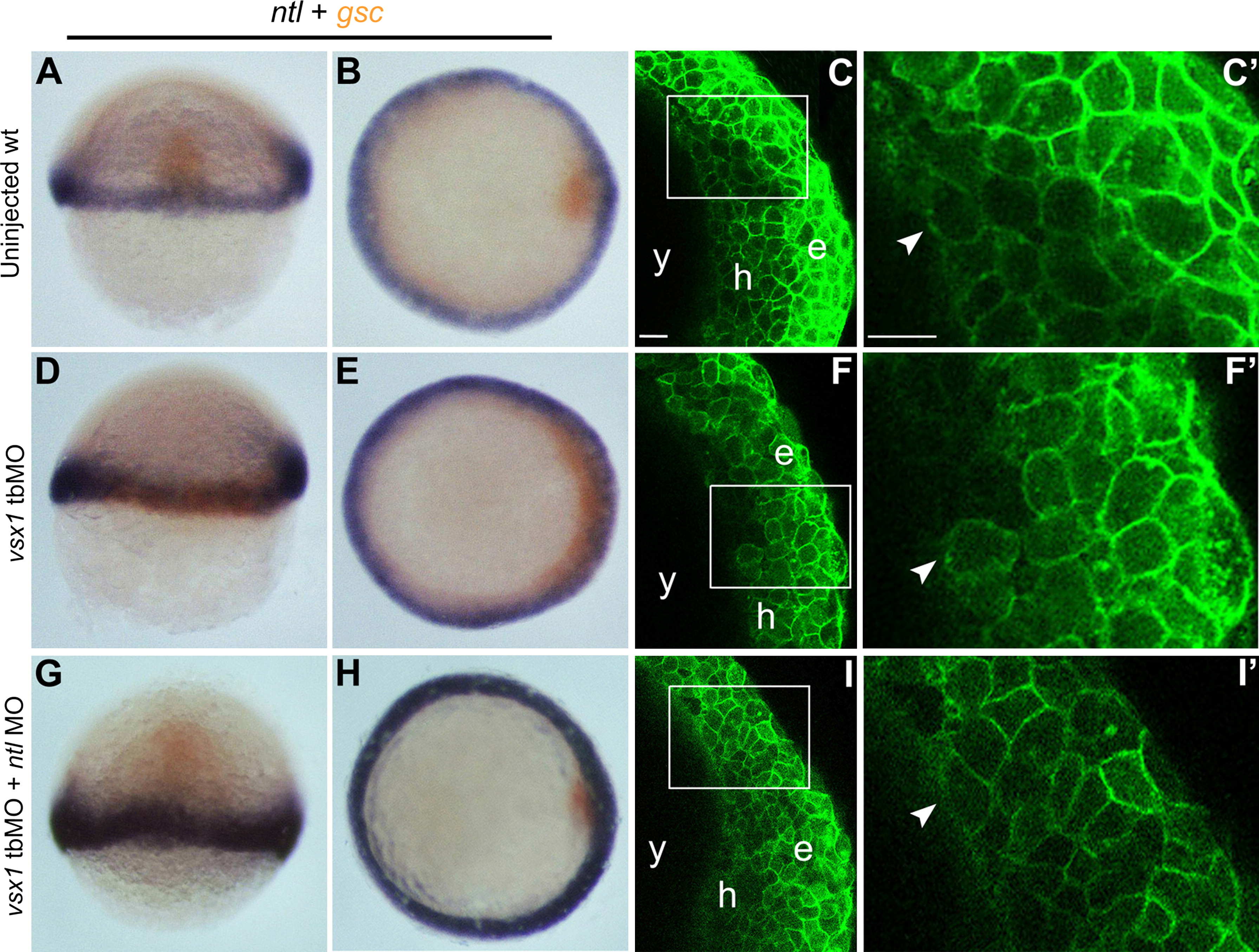
Maternal Vsx1 is essential for PME formation and progression. (A, B, D, E G, H) whole-mount in situ hybridization analysis of ntl (purple) and gsc (brown) expression in wild-type, vsx1 tbMO injected, vsx1 tbMO and ntl MO coinjected embryos at 40% epiboly. (C-C′, F-F′, I-I′) Cellular morphology and position of the anterior mesendoderm cells in wild-type, vsx1 tbMO injected, vsx1 tbMO and ntl MO coinjected embryos at 60% epiboly, observed by a confocal microscope. C′, F′ and I′ are the magnified image of C, F and I, respectively. The injected reagents are indicated at the left of each column. (A, D, G) Dorsal views with animal towards the top. (B, E, H) Animal views with dorsal towards the right. (C-C′, F-F′, I-I′) lateral views at the shield region with animal towards the top and dorsal towards the right. The arrowheads indicate the leading edge cells of the mesendoderm. e, h and y indicate epiblast, hypoblast and yolk sac, respectively. The bars represent 20 µm.
Reprinted from Developmental Biology, 394(2), Xu, X., He, Y., Sun, L., Ma, S., Luo, C., Maternal Vsx1 plays an essential role in regulating prechordal mesendoderm and forebrain formation in zebrafish, 264-76, Copyright (2014) with permission from Elsevier. Full text @ Dev. Biol.