
|
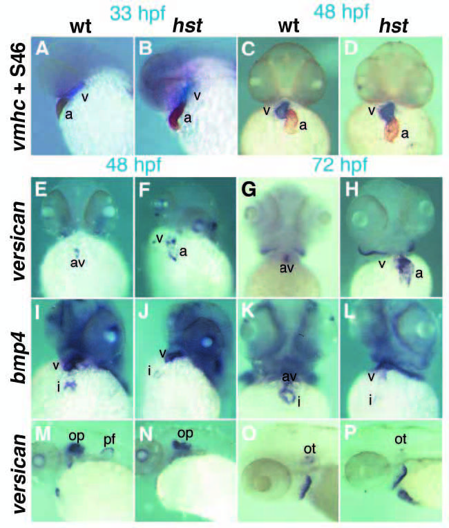
Fig. 7
Myocardial differentiation in hst mutant embryos. (A-D) In situ hybridization with the ventricle-specific marker vmhc (purple) followed by immunohistochemistry with the atrial-specific S46 antibody (brown). (A, wild type; B, hst) vmhc/S46 expression was normal at 33 hpf and (C, wild type; D, hst) at 48 hpf. (E-H) versican expression in the heart. At 33 hpf (not shown), wild-type and hst mutant embryos express versican broadly in the atrium and weakly in the ventricle. (E,F) 48 hpf; (G,H), 72 hpf; (E,G) wild-type embryos restrict versican expression to the AV boundary, but (F,H) hst mutant embryos fail to undergo this transition and continue to express versican predominantly in the atrium, with weak expression in the ventricle. (I-L) bmp4 expression in the heart. (I) Wild-type and (J) hst mutant embryos express bmp4 in the ventricle and inflow tract at 48 hpf. By 72 hpf (K) wild-type embryos restrict bmp4 to the AV junction, but (L) hst mutant embryos retain ventricle-enriched expression. (M-P) versican expression in the developing ear. At 48 hpf, (M) wildtype and (N) hst mutant embryos express versican broadly in the otic placode. By 72 hpf, (O) wild-type and (P) hst mutant embryos both restrict versican to the otoliths (Mowbray et al., 2001), suggesting the heart-specific defects in hst mutant embryos are not due to general developmental delay. a, atrium; av, atrioventricular boundary; i, inflow tract; op, otic placode; ot, otoliths; pf, pectoral fin bud; v, ventricle.