
Fig. 1
- ID
- ZDB-IMAGE-160503-4
- Genes
- Publication
- Valdivia et al., 2016 - Antagonism between Gdf6a and retinoic acid pathways controls timing of retinal neurogenesis and growth of the eye in zebrafish
- All Figures
- Figures for Valdivia et al., 2016
|
Fig. 1
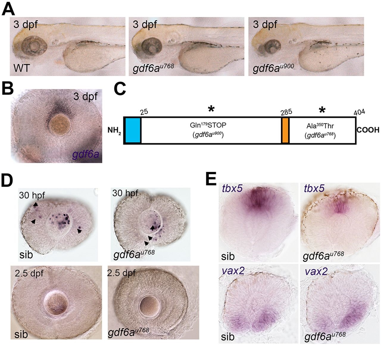
Small-eye phenotype in gdf6a mutants is independent of programmed cell death in developing retina. (A) Lateral views of wild-type and gdf6a mutants showing small eyes. (B) Lateral view of dorsal gdf6a expression (purple) in wild-type retina. (C) Domain structure of Gdf6a protein indicating the N-terminal signal peptide (amino acids 1-25, light blue), the furin protease recognition site (amino acids 280-285, orange), and the premature stop codon of gdf6au900 and missense mutation (Ala350Thr) in gdf6au768 (asterisks). (D) Lateral views of eyes of siblings (sib) and gdf6a mutants. Similar levels of TUNEL+ apoptotic cells (purple) are evident in the gdf6au768 and sibling eyes at 30hpf; most apoptotic cells are in the lenses (arrows) and only few in the retinae (arrowheads). TUNEL+ cells are undetectable in eyes of either genotype by 2.5dpf (lower panels). (E) Lateral views of whole-mount eyes of 24hpf gdf6au768 and sibling embryos showing expression (purple) of markers for dorsal (tbx5a) and ventral (vax2) retinal character.