
Fig. 5
- ID
- ZDB-IMAGE-160315-5
- Genes
- Publication
- Samuel et al., 2016 - Six3 regulates optic nerve development via multiple mechanisms
- All Figures
- Figures for Samuel et al., 2016
|
Fig. 5
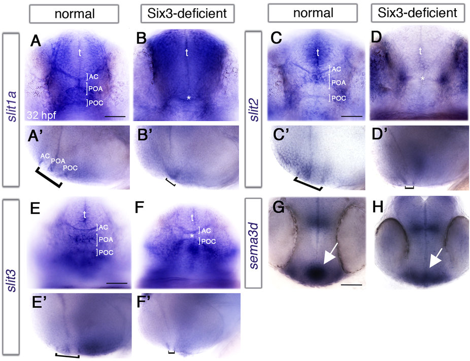
Abnormal midline signalling in Six3-deficient embryos.
(A-H) Expression of signalling molecules in anterior diencephalic midline of normal (A,C,E,G) and Six3-deficient (B,D,F,H) 32 hpf embryos. (A-F) frontal views, (A′-F′) the same embryos as in (A-F) respectively, lateral views. (A,A′,B,B′) slit1a expression. Asterisk in (B) marks an apparently single commissure. (C,C′,D,D′) slit2 expression. Asterisk in (D) marks what appears as a single commissure. (E,E′,F,F′) slit3 expression. Asterisk in F marks the reduced POA. Brackets in all lateral views mark the POA flanked by AC and POC. (G,H) sema3d expression. Arrows point at the POA. AC, anterior commissure; POA, preoptic area; POC, post-optic commissure; t, telencephalon. (G,H) are dorsal views, anterior down. Anterior is to the left in lateral views. Scale bars are 50 µm.