
Fig. 6
- ID
- ZDB-IMAGE-160226-56
- Genes
- Publication
- Gonsar et al., 2016 - Temporal and spatial requirements for Nodal-induced anterior mesendoderm and mesoderm in anterior neurulation
- All Figures
- Figures for Gonsar et al., 2016
|
Fig. 6
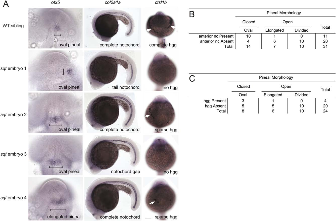
Positive correlation between neural tube closure and the presence of prechordal plate-derived hatching glands and axial mesoderm-derived anterior notochord. Embryos were raised to ~1 dpf and processed in tandem for otx5 in the pineal anlage, col2a1 in the notochord, and cstl1b in the hatching gland cells (hgg). (A) In WT embryos, the pineal organ is oval indicating a closed neural tube, the notochord spans the complete anterior–posterior extent of the trunk, and the hatching glands make a “necklace” around the anterior of the head. sqt embryos have a range of neural tube, notochord, and hgg defects as indicated. Note that, at this stage, col2a1 is expressed in three adjacent lines of cells, the floor plate (dorsal to the notochord), the hypochord (ventral to the notochord), and the notochord. The gaps in staining in sqt embryo 1 correspond to loss of all three tissues. The narrow region of staining in sqt embryo 3 just above the hind yolk corresponds to a place where the floor plate and hypochord cells persist, but the wider notochord cells are missing. The neural tube was scored as open if the pineal organ was at least twice as wide along its left–right axis (brackets) compared to its anterior–posterior axis. Images in the same row are different views of the same embryo. First column: dorsal views with anterior to the top. Second column: lateral views with anterior to the left and dorsal to the top. Third column: frontal views with anterior to the top. White arrows point to hatching glands. Scale bars: 100 µm. (B, C) Complete set of quantitative data.