
Fig. 2
- ID
- ZDB-IMAGE-160203-7
- Genes
- Publication
- Samsa et al., 2015 - Cardiac contraction activates endocardial Notch signaling to modulate chamber maturation in zebrafish
- All Figures
- Figures for Samsa et al., 2015
|
Fig. 2
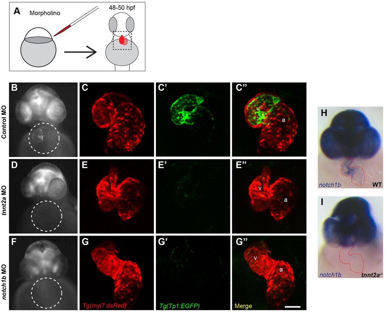
Cardiac contraction is required for endocardial Notch activation and notch1b transcription. (A) Schematic diagram of morpholino (MO) gene knockdown experiment, in which double transgenic Tg(Tp1:EGFP); Tg(myl7:dsRed) embryos were injected with (B,C) control, (D,E) tnnt2a or (F,G) notch1b morpholinos and imaged at 48-50hpf. (B,D,F) Representative whole-mount images of Notch reporter with cardiac regions highlighted by circles. (C,E,G) Confocal maximal intensity projections of the hearts shown in (B,D,F) with cardiomyocytes labeled in red, (C′,E′,G′) Notch reporters in green and (C′′,E′′,G′′) colocalized signal in yellow. Minimal colocalization indicates that Notch activation is in endocardial cells. (H,I) Whole-mount notch1b riboprobe hybridization in (H) control and (I) tnnt2a-/- embryos, with the heart outlined in red. Scale bar: 50µm. a, atrium; v, ventricle.