
IMAGE
Fig. 6
- ID
- ZDB-IMAGE-150615-6
- Genes
- Antibodies
- Publication
- Krock et al., 2014 - The Par-PrkC Polarity Complex Is Required for Cilia Growth in Zebrafish Photoreceptors
- All Figures
- Figures for Krock et al., 2014
Image
|
Figure Caption
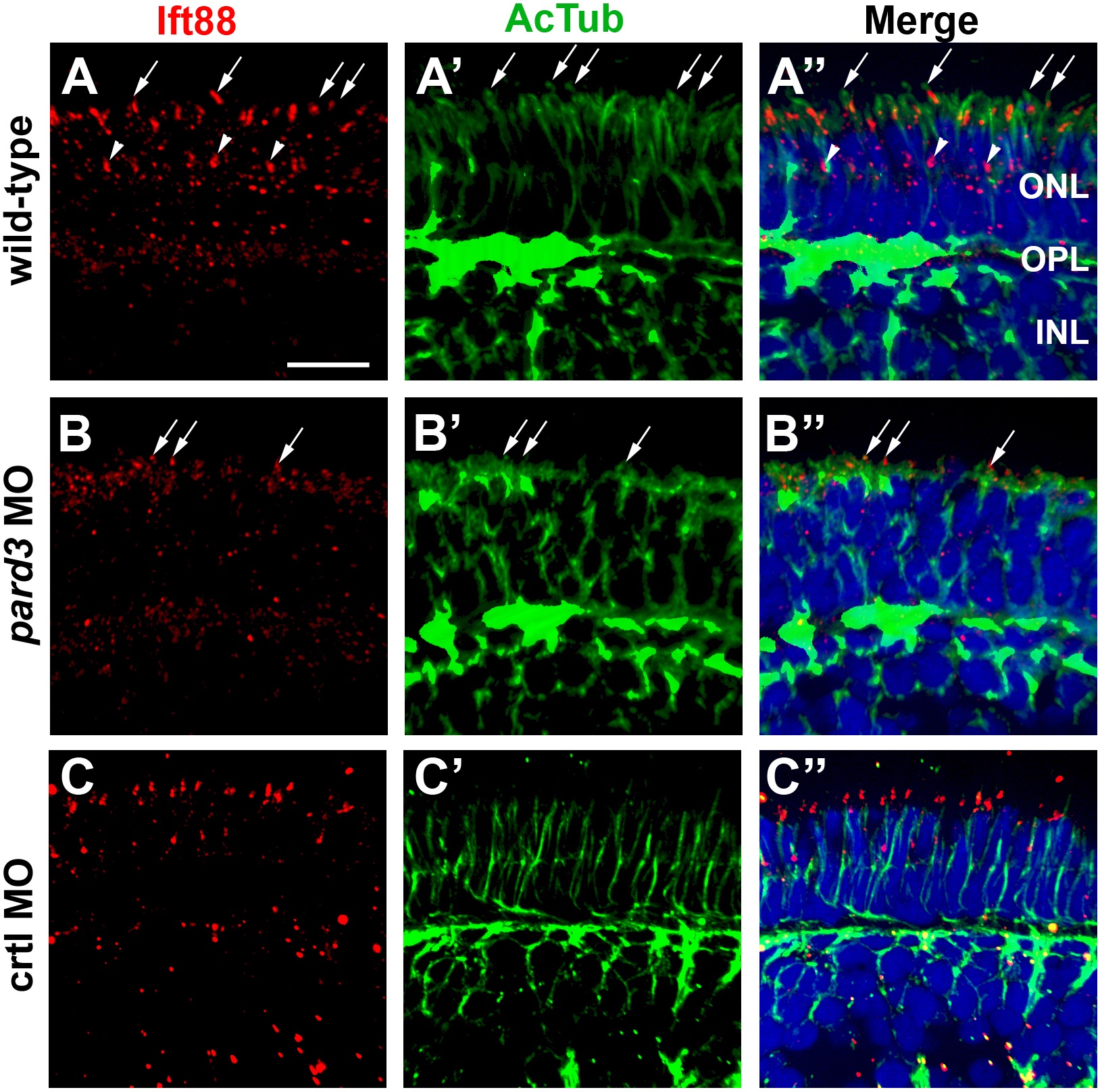
Fig. 6 Connecting cilia are reduced in pard3 morphants. (A–C) Transverse cryosections were stained for Ift88 (left, red) and acetylated tubulin (middle, green) to label cilia in 5 dpf retinas. Merged images are shown in right panels. Ift88 localizes to the connecting cilia emanating from the apical surface of photoreceptors (arrows). Staining is also seen in connecting cilia of UV cones (arrowheads), which are tiered below the other cones. Sections were also counterstained with DAPI (blue). ONL = outer nuclear layer; OPL = outer plexiform layer; INL = inner nuclear layer. Scale bar = 10 μm.
Figure Data
Acknowledgments
This image is the copyrighted work of the attributed author or publisher, and
ZFIN has permission only to display this image to its users.
Additional permissions should be obtained from the applicable author or publisher of the image.
Full text @ PLoS One