
Fig. 9
- ID
- ZDB-IMAGE-150602-31
- Genes
- Publication
- Liu et al., 2015 - Differential expression of protocadherin-19, protocadherin-17, and cadherin-6 in adult zebrafish brain
- All Figures
- Figures for Liu et al., 2015
|
Fig. 9
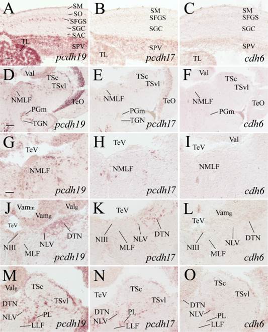
Expression of pcdh19, pcdh17, and cdh6 in the optic tectum, dorsal tegmentum, isthmus, and cerebellar valvula. A–C: Higher magnifications of adjacent sections of the medial optic tectum from images shown in the top panels in Fig. 7. D–F: Low magnified views of dorsal tegmental region of adjacent sections at a level shown in Fig. 1. G–I: Higher magnifications of the nucleus of medial longitudinal fascicle (NMLF) of their respective images in D–F. J–O: Magnified views of the dorsal tegmentum and isthmus from sections posterior (120–150 µm) to D–F, with J–L showing the medial region, while M–O showing the lateral region. See list for abbreviations. Scale bar = 100 µm for D–F, 50 µm for the remaining panels (G–O have the same magnification).