
Fig. 5
- ID
- ZDB-IMAGE-150421-9
- Genes
- Publication
- Yuan et al., 2015 - Intraciliary Calcium Oscillations Initiate Vertebrate Left-Right Asymmetry
- All Figures
- Figures for Yuan et al., 2015
|
Fig. 5
Intraciliary Calcium Is Essential for Vertebrate LR Development
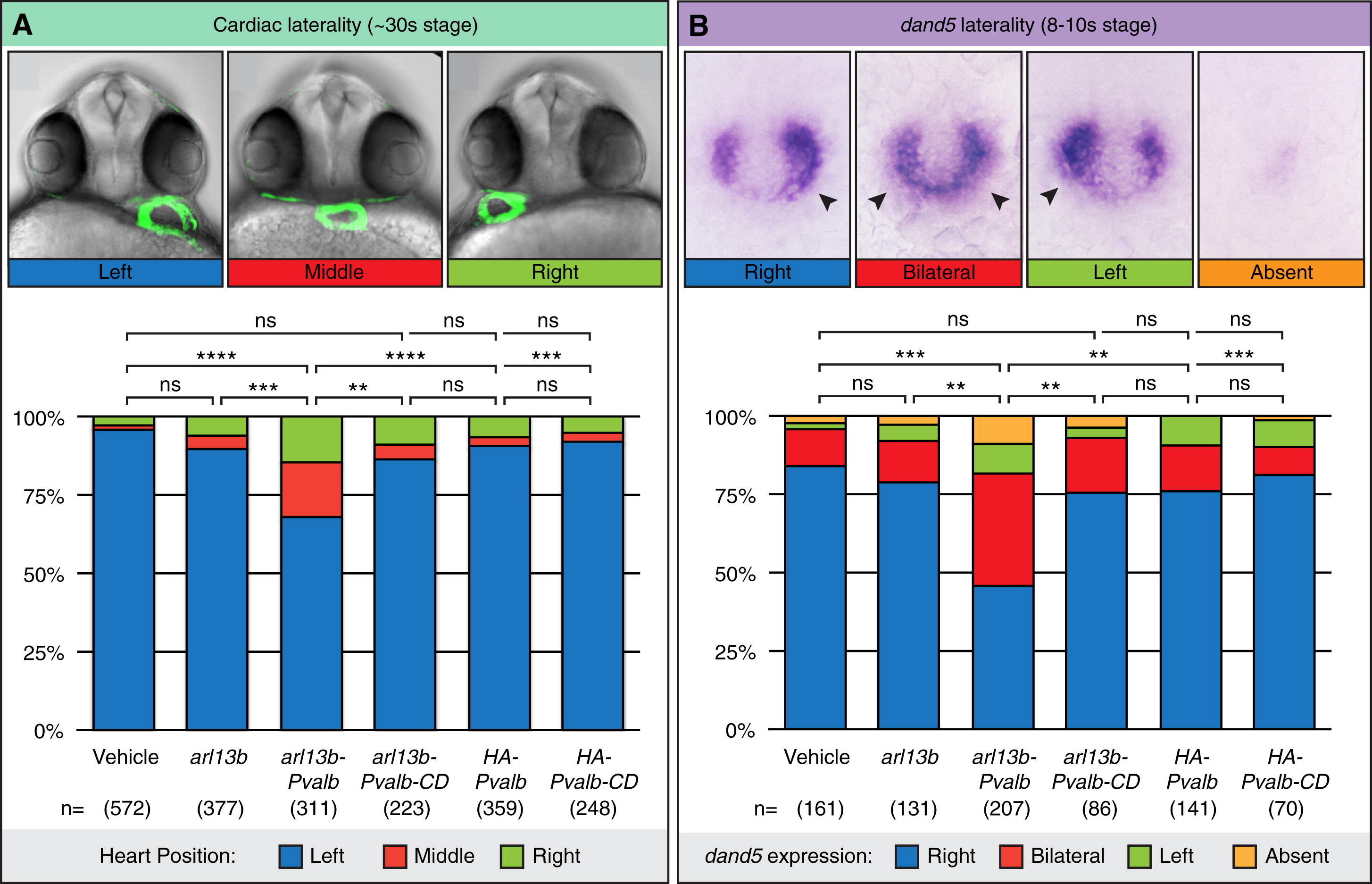
(A) Representative bright-field and fluorescent images showing the heart position in zebrafish embryos at <30-somite stage highlighted by a GFP transgene driven by the promoter of cardiac myosin light chain 2. The embryo is seen from the ventral side, showing normal left (blue) and abnormal middle (red) and right (green) heart loops. The graph shows the distribution of heart position in response to expression of parvalbumin and mutant parvalbumin (PVALB-CD) targeted to cilia or cytoplasm of wild-type embryos. Data shown are pooled from nine total independent experiments.
(B) Representative images of whole-mount in situ hybridization for dand5 (charon) expression in the LRO of zebrafish embryos at the eight- to ten-somite stage. The embryo is seen from the dorsal side, showing normal right-sided (blue) expression and abnormal bilateral (red), left-sided (green), or absent (orange) expression. Arrowheads indicate handedness of dand5 expression across the left-right axis. The graph shows the distribution of dand5 in response to expression of PVALB and PVALB-CD targeted to cilia or cytoplasm.
Data shown are pooled from three independent experiments. Statistical comparison was analyzed by one-way ANOVA with Tukey’s multiple comparison test; p < 0.005, p < 0.0005, p < 0.0001; ns: p e 0.05. n, total number of embryos analyzed for each experimental condition (in parentheses). See also Figure S6.