
Fig. 6
- ID
- ZDB-IMAGE-150413-38
- Genes
- Publication
- Jackson et al., 2015 - The role of Sox6 in zebrafish muscle fiber type specification
- All Figures
- Figures for Jackson et al., 2015
|
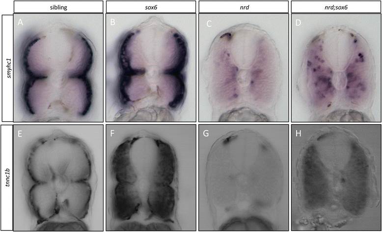
Fig. 6
Elimination ofsox6rescuestnnc1bandsmyhc1expression inprdm1anrdmutants. (A-D) Cross sections through the trunk region of embryos that were hybridized with the smyhc1 probe at 30 hours postfertilization (hpf): note that migration of the slow-twitch progenitors is disrupted in prdm1a mutants, and the expression of smyhc1 is dramatically downregulated (cf A and C); in the prdm1a;sox6 double mutant, smyhc1 expression is restored in the adaxial cells, though their migration still appears disrupted (cf. C and D). (E-H) Similar cross sections of wild-type and mutant embryos hybridized with the tnnc1b probe. Expression of tnnc1b is completely lost prdm1a mutants (cf E and G); expression is restored throughout the myotomes in the prdm1a;sox6 double mutants, though at lower levels than in the sox6 single mutant (cf. F and H) are slow specific genes, which are expressed in the superficial slow-twitch fibers in wild-type embryos at 30 hpf (A and B).