
Fig. 5
- ID
- ZDB-IMAGE-150330-11
- Genes
- Publication
- Marín-Juez et al., 2015 - GLUT2-mediated glucose uptake and availability are required for embryonic brain development in zebrafish
- All Figures
- Figures for Marín-Juez et al., 2015
|
Fig. 5
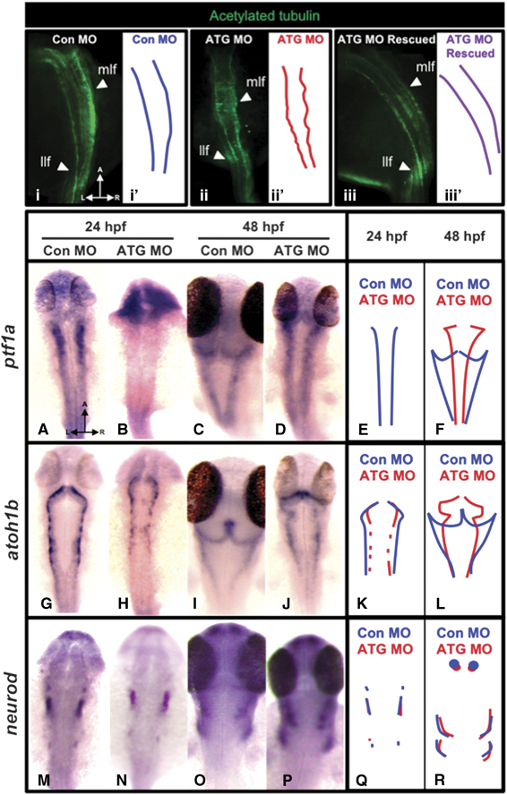
glut2 abrogation causes hindbrain disorganization and affects the expression of cerebellar proneural genes. To study the consequences of glut2 knockdown in the hindbrain structure we performed immunostaining using an antibody against acetylated tubulin in con MO, ATG MO, and ATG MO+rat GLUT2 mRNA rescued embryos at 24hpf (i, ii, iii). At this stage, morphant embryos showed disorganized axon tracts. Rescued embryos showed a hindbrain structure similar to control injected embryos. Lateral longitudinal fascicles (llf); medial longitudinal fascicles (mlf). To further study the consequences of glut2 abrogation in the neural progenitor cells we performed ISH for the proneural genes ptf1a (A–D), atoh1b (G–J), neurod (M–P) in control injected embryos at 24hpf (A, G, and M), and 48hpf (C, I, and O), and in ATG morphants at 24hpf (B, H, and N) and 48hpf (D, J, and P). To better illustrate the effects caused by the abrogation of glut2, immunostained medial longitudinal fascicles have been outlined (i′, ii′, iii′). The expression patterns observed by ISH of the proneural genes are represented with diagrams overlapping the expression patterns in control and ATG morphants at 24hpf and 48hpf of ptf1a (E and F), atoh1b (K and L), and neurod (Q and R). A, anterior; L, left; R, right.