
Fig. 5
- ID
- ZDB-IMAGE-150324-5
- Genes
- Publication
- Sawamiphak et al., 2014 - Interferon Gamma Signaling Positively Regulates Hematopoietic Stem Cell Emergence
- All Figures
- Figures for Sawamiphak et al., 2014
|
Fig. 5
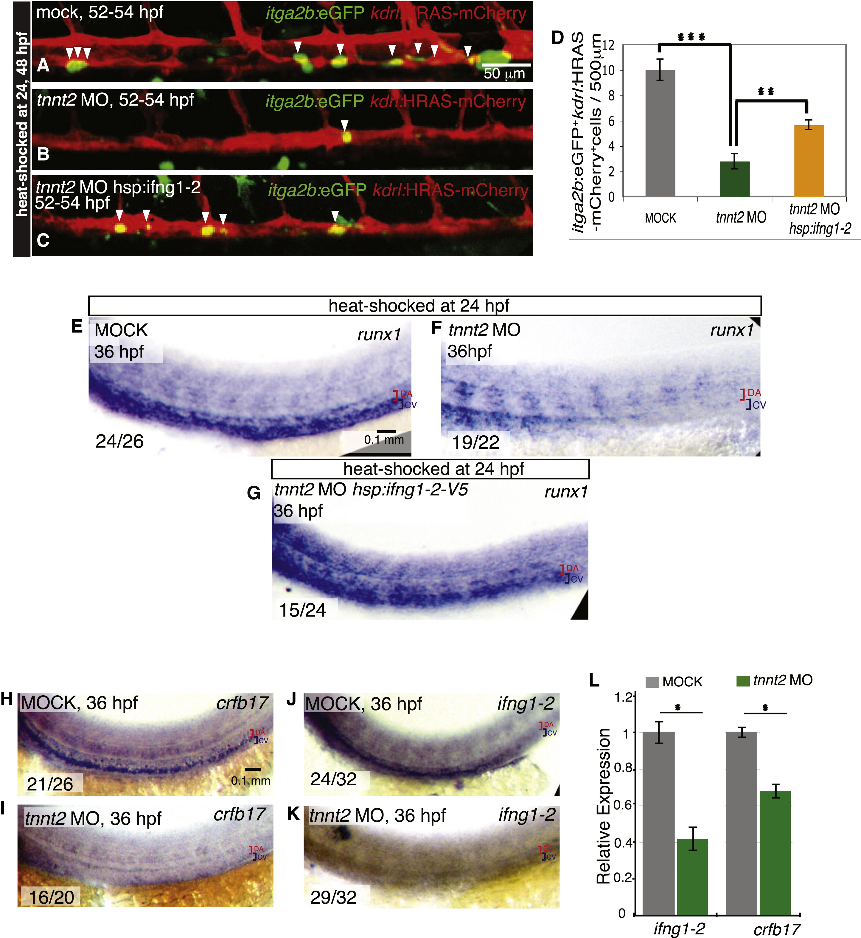
Reduced Ifng1-2 Signaling Is Partially Responsible for the HSC Developmental Defects Caused by the Lack of Blood Flow
(A–D) ifng1-2 overexpression partly restores HSCs in embryos with no circulation. itga2b:EGFP+ (green) kdrl:HRAS-mCherry+ (red) HSCs (white arrowheads) are shown in mock-injected controls (A) and tnnt2 MO-injected embryos without (B) and with (C) hsp:ifng1-2 transgene. All embryos were heat shocked at 24 and 48 hpf and imaged at 52–54 hpf. (D) HSC numbers per 500 µm aortic length are shown as means ± SEM, n = 12–20 embryos, p d 0.01, p d 0.001.
(E–G) Loss of runx1 expression from the lack of blood flow is rescued by ifng1-2 overexpression. runx1 expression is shown in mock-injected controls (E) and tnnt2 MO-injected embryos without (F) and with (G) the hsp:ifng1-2 transgene. All embryos were heat-shocked at 24 hpf and harvested at 36 hpf.
(H–K) crfb17 and ifng1-2 expression depends on blood flow. crfb17 (H and I) and ifng1-2 (J and K) expression is shown in mock-injected (H and J) and tnnt2 MO-injected (I and K) 36 hpf embryos. The number of embryos showing the representative phenotype per total number of embryos analyzed is indicated in the lower left corner. Red brackets identify the dorsal aorta (DA), and blue brackets identify the cardinal vein (CV). All images are lateral views, dorsal up and anterior to the left. qPCR assay of ifng1-2 and crfb17 expression at 36 hpf in tnnt2 MO-injected embryos compared with mock-injected siblings, n = 3, 30 embryos per sample, p d 0.05.
(L) qPCR assay of ifng1-2 and crfb17 expression at 36 hpf in tnnt2 MO-injected embryos compared with mock-injected siblings, n = 3, 30 embryos per sample, p d 0.05.
Reprinted from Developmental Cell, 31, Sawamiphak, S., Kontarakis, Z., Stainier, D.Y., Interferon Gamma Signaling Positively Regulates Hematopoietic Stem Cell Emergence, 640-653, Copyright (2014) with permission from Elsevier. Full text @ Dev. Cell