
Fig. 10
- ID
- ZDB-IMAGE-140306-10
- Genes
- Publication
- Reifers et al., 1998 - Fgf8 is mutated in zebrafish acerebellar (ace) mutants and is required for maintenance of midbrain-hindbrain boundary development and somitogenesis
- All Figures
- Figures for Reifers et al., 1998
|
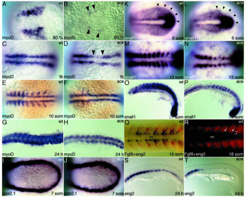
Fig. 10 Fgf8 is involved in mesoderm and somite patterning. (A,B) Expression of myoD is strongly reduced in adaxial mesoderm of acerebellar embryos at 80% epiboly (arrowheads point to remnants of expression). (C,D) At tailbud stage, myoD expression in adaxial mesoderm is interrupted in acerebellar (arrowheads). (E,F) myoD staining in the somitic mesoderm is strongly reduced in mutants at the 10- somite stage. (G,H) At 24 hours, the expression of myoD is weak in the smaller and less-well-differentiated somites of acerebellar embryos. (I,J) No obvious difference could be detected between wild-type and acerebellar embryos in formation of intermediate mesoderm, shown here with Pax2.1 staining at the 7-somite stage. (K,L) Expression of snail1 is reduced in acerebellar embryos in the region around the tailbud at 6- somite stage (arrowheads point to the wild-type border of expression). (M,N) At the 13-somite stage and (O,P) 20-somite stage, snail1 transcripts are strongly reduced in the somites of mutant embryos. (Q,R) Dorsal view of wild-type embryo stained for eng2 (blue) and Fgf8 (red, fluorescent) showing partial overlap of these expression domains at an early stage of somite development (arrows). Note the restriction of Fgf8 expression to anterolateral cells of the somites over time (anterior is to the left; brackets depict adaxial cells; nc, notochord). (S,T) Muscle pioneers are reduced in acerebellar embryos, as shown here for 24 hours embryos with Eng2 staining.