Fig. 2
Fig. 2
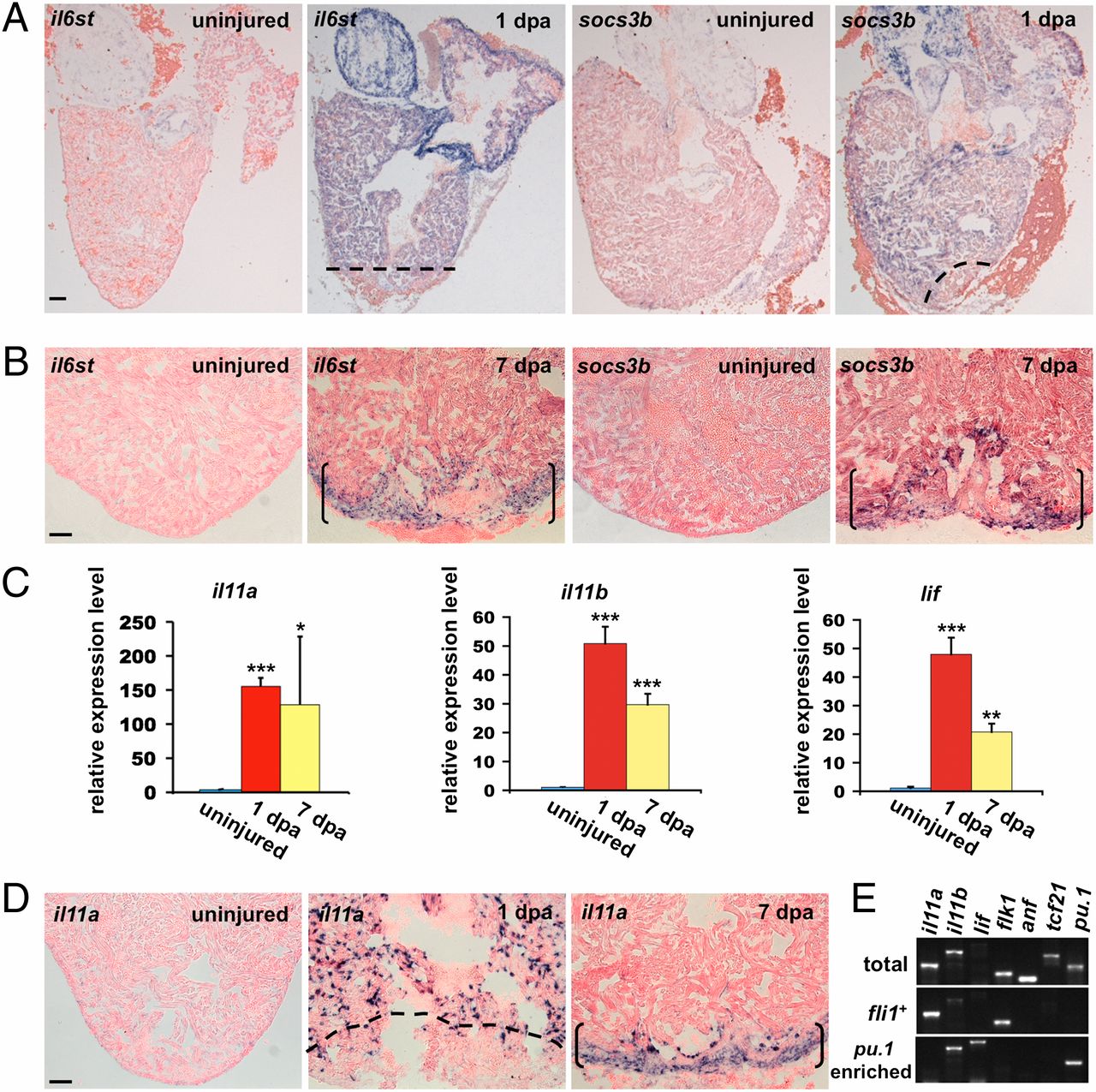
Induction of Jak1/Stat3 pathway members and ligands after cardiac injury. (A and B) In situ hybridization revealing Jak1/Stat3 pathway members (shown here are il6st and socs3b) induced in an organ-wide manner at 1 dpa (A), and then restricted to the injury site at 7 dpa (B). Dashed lines indicate approximate amputation plane. Brackets indicate injury site. (C) qPCR from ventricular RNA samples revealed induction of il11a, il11b, and lif at 1 and 7 dpa. Data are mean ± SEM n = 3, *P < 0.05, **P < 0.01, ***P < 0.001, Student t test (unpaired, two-tailed). Expression levels were normalized to that of β-actin2, and further normalized to that of the uninjured sample. (D) il11a expression was induced in an organ-wide manner in endocardial cells at 1 dpa, and localized to the injury site at 7 dpa. Dashed line indicates approximate amputation plane. Brackets indicate injury site. (E) RT-PCR of samples from purified endocardial/endothelial (fli1:EGFP+) cells or pu.1-enriched cells at 1 dpa. il11a was detected in fli1:EGFP+ samples, similar to the Vegf receptor flk1. lif was detected in pu.1-enriched samples; it was undetectable when the same amount of total ventricular RNA was used for amplification (total). il11b was detected in pu.1-enriched samples and less so in fli1:EGFP+ samples. anf and tcf21 are markers for cardiomyocytes and epicardium, respectively, and were detected in total ventricular RNA samples. (Scale bars, 50 μm.)

