
Fig. S9
- ID
- ZDB-IMAGE-121030-21
- Genes
- Antibodies
- Publication
- Hinits et al., 2012 - Zebrafish Mef2ca and Mef2cb are essential for both first and second heart field cardiomyocyte differentiation
- All Figures
- Figures for Hinits et al., 2012
|
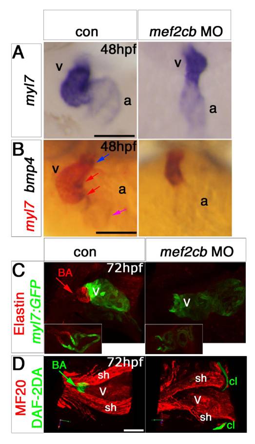
Fig. S9
Mef2cb morphants have a linear defective heart and loss of bulbus arteriosus.
In situ mRNA hybridisation for indicated genes (A and B, ventral view, anterior to top) and immunodetection of EGFP, MyHC (MF20), Elastin and DAF-2DA detection (C and D, lateral view, anterior to left) of hearts at indicated stage of mef2cb morphants and controls. A and B. Mef2cb morphants have a linear unlooped hearts and no obvious AV canal as marked by bmp4 expression (red arrows). C and D. Confocal stacks of control or mef2cb MO-injected Tg(myl7:EGFP) (E) or wild-type (F) embryos at 72 hpf. Hearts are unlooped and bulbus arteriosus markers (BA, arrows) are absent in mef2cb morphants. Insets in D show YZ sections at the OFT region, indicating to the area bordering myocardial and smooth muscle cells. sh, sternohyoideus; cl, cleithrum; v, ventricle; a, atrium IFT, inflow tract; OFT, outflow tract; AVC, atrioventricular canal. Scale = 100 μm, except for C and D= 50 μm.
Reprinted from Developmental Biology, 369(2), Hinits, Y., Pan, L., Walker, C., Dowd, J., Moens, C.B., and Hughes, S.M., Zebrafish Mef2ca and Mef2cb are essential for both first and second heart field cardiomyocyte differentiation, 199-210, Copyright (2012) with permission from Elsevier. Full text @ Dev. Biol.