
Fig. S8
- ID
- ZDB-IMAGE-121030-20
- Genes
- Antibodies
- Publication
- Hinits et al., 2012 - Zebrafish Mef2ca and Mef2cb are essential for both first and second heart field cardiomyocyte differentiation
- All Figures
- Figures for Hinits et al., 2012
|
Fig. S8
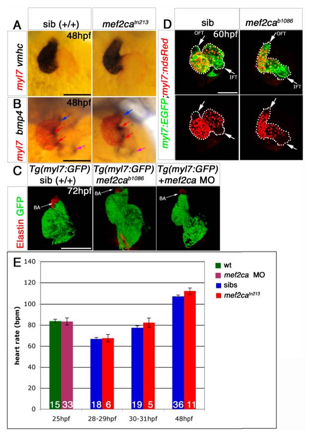
Mef2ca mutant has a normal heart.
A and B. Double mRNA in situ hybridisation for myl7 (red, A and B), vmhc (blue, A) and bmp4 (blue, B) in genotyped 48 hpf mef2ca mutant embryos and their siblings shown in ventral view, anterior to top. Mutants have normal looping and gene expression is no different than siblings. OFT, blue arrow, IFT, purple arrow, AVC, red arrows. C. Confocal stacks of 72 hpf mef2cab1086;Tg(myl7:GFP) mutants and their siblings and of Tg(myl7:GFP) injected with mef2ca MO immunostained for Elastin showing no difference between morphants, mutants and siblings. D. GFP+dsRED- CMs can only be found in OFT and IFT of the heart (dotted line) of mef2cab1086;Tg(myl7:GFP;myl7;ndsRed) mutants and their siblings at 60 hpf. E. Heart rate (bpm) of control wild type, mef2camorphants, mef2catn213 and their siblings at 25-48 hpf. Embryos were grown in 24-well plates and genotype was determined at 5 dpf according to jaw phenotype (see Piotrowski et al., 1996). No significant differences according to t-test statistics were found at any stage between morphants/mutants and control/siblings. Number of embryos per condition is shown on columns. Variation in heart rate between experiments is due to variation in temperature. Scale = 100 μm.
Reprinted from Developmental Biology, 369(2), Hinits, Y., Pan, L., Walker, C., Dowd, J., Moens, C.B., and Hughes, S.M., Zebrafish Mef2ca and Mef2cb are essential for both first and second heart field cardiomyocyte differentiation, 199-210, Copyright (2012) with permission from Elsevier. Full text @ Dev. Biol.