
Fig. 3
- ID
- ZDB-IMAGE-120810-34
- Genes
- Publication
- Johnson et al., 2012 - Scube activity is necessary for Hedgehog signal transduction in vivo
- All Figures
- Figures for Johnson et al., 2012
|
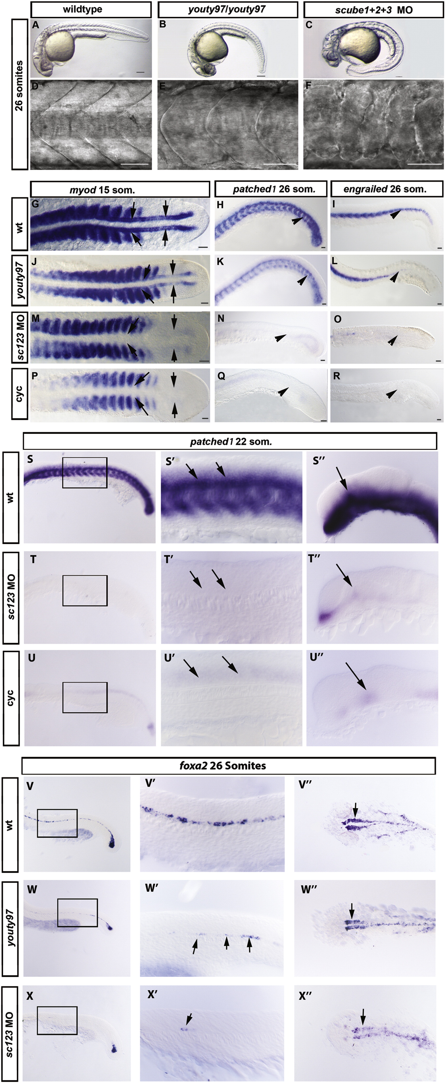
Fig. 3 scube1 and scube3 morpholinos enhance the phenotype of youty97 mutant embryos. (A–F). Somite shape in live 30 hpf embryos, lateral view anterior to left. (A, D) The characteristic chevron shape of the wildtype zebrafish somite became rounded or ‘U’ shaped in the youty97 mutant (B, E). (C, F) Morpholino knockdown of all 3 scube genes enhanced the youty97 mutant phenotype, a severe loss of the chevron shape resulted in ‘blocky’ shaped somites. Scale Bars: A–C 150 μm, D–F 100 μm. (G–R). In situ hybridization revealed Hh target gene expression (myod, patched1 (ptc1), and engrailed (eng)), decreased dramatically in scube triple MO embryos. (G) myod is expressed in somites and adaxial cells of wildtype embryos, (J) adaxial expression of myod is reduced in youty97 homozygous mutants, (M) adaxial myod expression is absent from scube triple MO embryo. (P) Adaxial myod expression is also absent from cyclopamine treated embryos. (G, J, M, P) Arrows indicate location of adaxial cells. (H) ptc expression in ventral neural tube and somites in wildtype embryos. (K) Reduced ptc expression in posterior somites of youty97 embryo, (N) somitic expression of ptc is absent from scube triple MO and (Q) cyclopamine treated embryos. (H, K, N, Q) Arrowheads highlight regions of altered gene expression. (I) In wildtype embryos eng is expressed in the muscle pioneer cells and a subset of fast muscle fibers, (L) eng expression is reduced in the posterior of youty97 mutant embryos, (O) in scube triple MO embryos eng expression is severely reduced with only small patches of eng positive cells visible. (R) Cyclopamine treated embryos display the most severe loss of eng staining. (I, L, O, R) Arrowheads highlight regions of altered gene expression. (G, J, M, P) 15 somite stage, dorsal view, (H, K, N, Q) 22 somite stage, lateral view anterior to left, (I, L, O, R) 26 somite stage, lateral view, anterior to left. Scale bars: 50 μm. (S–U′′) ptc1 expression is reduced in neural tissue of scube triple MO embryos. In wildtype, uninjected 22 somite embryos (S–S′′), ptc1 expression is prominent in the ventral neural tube of the trunk (S and arrows in S′) as well as the somites, where its expression is known to be induced by HH signaling from the midline. In the brain, ptc1 is highly expressed within the diencephalon (S′′, arrow), an expression domain known to be induced by HH signaling from the underlying zona limitans intrathalamica. In both scube triple MO embryos (T–T′′) and in cyclopamine treated embryos (U–U′′) expression of ptc1 in both these tissues is severely reduced or absent. (V–X′′) Expression of the ventral neural marker foxa2 is severely reduced in scube triple MO embryos. In wildtype embryos at 26 somites foxa2 is expressed in floor plate (V, V′ both medial and lateral cells) and in two wings of expression in the ventral diencephalon (arrow, V′). In homozygous youty97 mutants both these regions of expression are reduced (Fig. 3W–W′′), and in scube triple MO embryos this reduced expression is greatly enhanced (X–X3). (S–X′′) Within each row, the same letter indicates a different region of the same embryo.′ Add –(S–X′′) anterior to left, (S–U′′) 22 somites, lateral view (V,V′, W, W′, X, X′) 26 somites, lateral view, (V′′, W′′, X′′) dorsal view.
Reprinted from Developmental Biology, 368(2), Johnson, J.L., Hall, T.E., Dyson, J.M., Sonntag, C., Ayers, K., Berger, S., Gautier, P., Mitchell, C., Hollway, G.E., and Currie, P.D., Scube activity is necessary for Hedgehog signal transduction in vivo, 193-202, Copyright (2012) with permission from Elsevier. Full text @ Dev. Biol.