
Fig. 3
- ID
- ZDB-IMAGE-120611-13
- Genes
- Antibodies
- Publication
- Manfroid et al., 2012 - Zebrafish sox9b is crucial for hepatopancreatic duct development and pancreatic endocrine cell regeneration
- All Figures
- Figures for Manfroid et al., 2012
|
Fig. 3
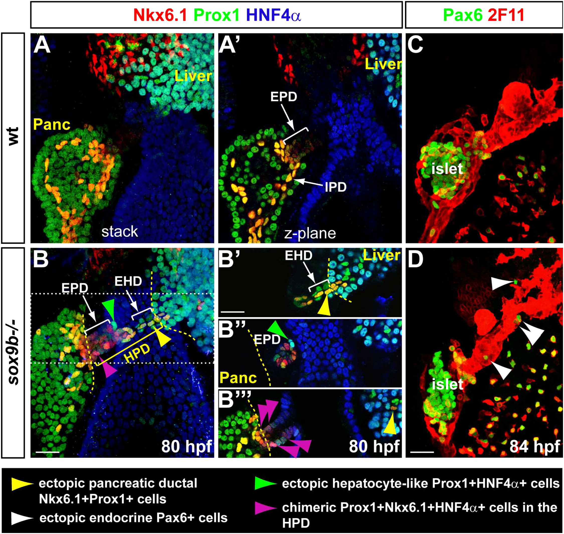
Misdifferentiation of the HPD system in sox9b mutants. (A, B) 3D-rendering (stack) at 80 hpf of the liver, pancreas and HPD system immunolabelled with Prox1, HNF4α and the pancreatic ductal marker Nkx6.1. (A2, B2–B32) Z-planes through the same larvae as in A and B. Prox1 with HNF4α label hepatocytes while Prox1 together with Nkx6.1 strongly labels the IPD. In sox9b mutants (B and B2–B32), the HPD system becomes labelled with the three markers and display different ectopic cell types (see the colour code at the bottom). Dotted lines delimitate organs and ducts boundaries. (C, D) 3-D rendering of the HPD system at 84 hpf labelled with the ductal marker 2F11 and the endocrine marker Pax6. Ectopic endocrine cells along the HPD and in the liver are indicated by white arrowheads. EHD, extrahepatic duct; EPD, extrapancreatic duct; HPD, hepatopancreatic ductal system; IPD, intrapancreatic ducts. Scale bar=20 μm.
Reprinted from Developmental Biology, 366(2), Manfroid, I., Ghaye, A., Naye, F., Detry, N., Palm, S., Pan, L., Ma, T.P., Huang, W., Rovira, M., Martial, J.A., Parsons, M.J., Moens, C.B., Voz, M.L., and Peers, B., Zebrafish sox9b is crucial for hepatopancreatic duct development and pancreatic endocrine cell regeneration, 268-278, Copyright (2012) with permission from Elsevier. Full text @ Dev. Biol.