
Fig. 1
- ID
- ZDB-IMAGE-111020-17
- Genes
- Publication
- He et al., 2011 - miR-196 regulates axial patterning and pectoral appendage initiation
- All Figures
- Figures for He et al., 2011
|
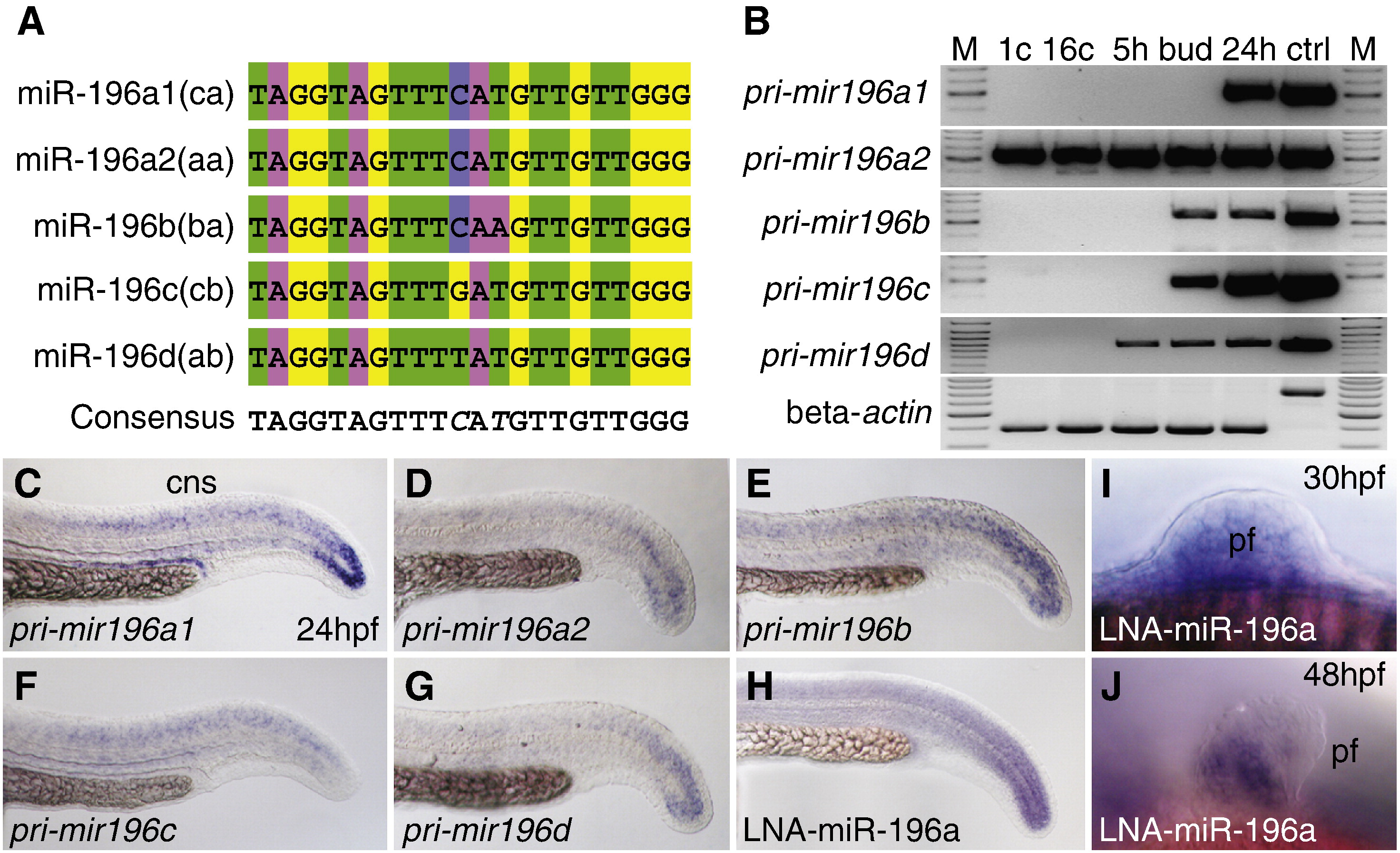
Fig. 1 Sequence and expression of mir196 genes. (A) Alignment of mature mir196 encoded by five mir196 genes. (B) Expression of mir196 paralogs studied by RT-PCR. Beta-actin (bactin1) was used as control for contaminating genomic DNA (ctrl lane). M, size marker; 1c, 1 cell stage; 16c, 16 cell stage; 5 h, 5 hpf (hours post-fertilization); bud, bud stage, about 10 hpf; 24 h, 24 hpf; ctrl, genomic DNA control. (C–H) Whole mount in situ hybridization for mir196 primary transcripts showed expression in the tail bud and neural tube. (H) Linked nucleic acid (LNA) probe for miR-196a showed an expression pattern similar to the primary transcript. (I, J) LNA probes for mir196a in the pectoral fin bud at 30 and 48 hpf. cns, central nerve system; pf, pectoral fin.
Reprinted from Developmental Biology, 357(2), He, X., Yan, Y.L., Eberhart, J.K., Herpin, A., Wagner, T.U., Schartl, M., and Postlethwait, J.H., miR-196 regulates axial patterning and pectoral appendage initiation, 463-77, Copyright (2011) with permission from Elsevier. Full text @ Dev. Biol.