
Fig. 5
- ID
- ZDB-IMAGE-110812-26
- Genes
- Antibodies
- Publication
- Naganawa et al., 2011 - Developmental transition of touch response from slow muscle-mediated coilings to fast muscle-mediated burst swimming in zebrafish
- All Figures
- Figures for Naganawa et al., 2011
|
Fig. 5
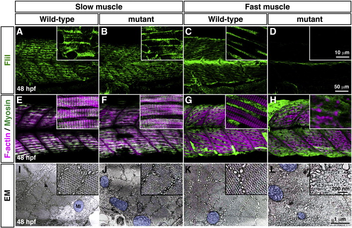
Actin–myosin fibers are disorganized in flii fast muscles at 48 hpf. (A–D) The expression of FliI protein was assayed with anti-FliI at 48 hpf. FliI protein was expressed in wild-type slow muscle (A), mutant slow muscle (B) and wild-type fast muscle (C) but not in mutant fast muscle (D). (E–H) Double labeling with phalloidin (F-actin) and anti-myosin (slow muscle: F59; fast muscle: MF20) at 48 hpf. Co-labeling of F-actin and slow myosin was seen in wild-type (E) and mutant (F). F-actin was organized in wild-type fast muscle (G) but not in mutant fast muscle (H). (I–L) Electron micrographs of cross-sections of muscle at 48 hpf. Actin and myosin bundles were organized in wild-type slow muscle (I), mutant slow muscle (J) and wild-type fast muscle (K) but not in mutant fast muscle (L). Mitochondria are shaded blue.
Reprinted from Developmental Biology, 355(2), Naganawa, Y., and Hirata, H., Developmental transition of touch response from slow muscle-mediated coilings to fast muscle-mediated burst swimming in zebrafish, 194-204, Copyright (2011) with permission from Elsevier. Full text @ Dev. Biol.